Liquid ready-to-use prothrombin time detection reagent

A technology for prothrombin time and detection reagents, which is applied in the field of biomedical diagnosis, can solve the problems of large batch-to-batch variation and freeze-dried powder preparation bottle-to-bottle variation, and achieves good stability, easy quality control, and high sensitivity. Effect

Inactive Publication Date: 2017-11-17
NINGBO ACCUTECH BIOSCI LTD
View PDF7 Cites 12 Cited by
  • Summary
  • Abstract
  • Description
  • Claims
  • Application Information

AI Technical Summary

Problems solved by technology

Because there will be differences when subpackaging, and the freeze-drying effect of each bottle is different during the freeze-drying process, and the concentration difference caused by the different amount of reconstitution agent added during the reconstitution process, all these differences make the difference between the freeze-dried powder preparation bottles Poor, large variation between batches

Method used


Image

  • Liquid ready-to-use prothrombin time detection reagent
  • Liquid ready-to-use prothrombin time detection reagent
  • Liquid ready-to-use prothrombin time detection reagent

Examples


Embodiment 1

[0018] The preparation of embodiment 1 prothrombin time detection reagent

[0019] (1) Lipidation of tissue factor

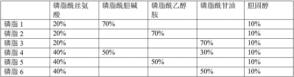
[0020] Take an appropriate amount of synthetic phospholipids by weighing respectively: phosphatidylserine, phosphatidylcholine, phosphatidylglycerol, cholesterol, and dissolve in the surfactant OGP solution in different proportions (accounting for the mass percentage of synthetic phospholipids). See Table 1 for details. The final concentration is 25mg / ml, mix until the synthetic phospholipids are completely dissolved, add 100μg / ml recombinant rabbit tissue factor, stir and incubate at room temperature for 2 hours, transfer the solution to a dialysis bag, change the dialysate every 4 hours, dialyze Overnight, remove the surfactant OGP. The obtained lipidated recombinant rabbit tissue factor solution was diluted with Hepes buffer to prepare a PT reagent containing 250 μg / ml synthetic phospholipids and 1 μg / ml recombinant rabbit tissue factor.

[0021] Table 1 Di...

Embodiment 2

[0035] The mensuration of embodiment 2 stability

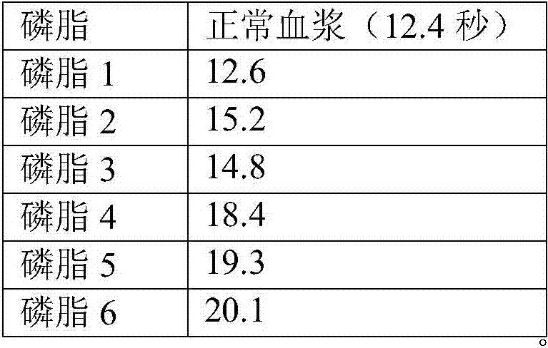
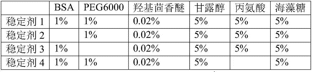
[0036] According to the method of Example 1, the PT detection reagent was prepared using phospholipid 1 and stabilizer 5. PT detection reagent contains 20mM, HEPES buffer, 200mM sodium chloride, 5% alanine, 5% trehalose, 1% BSA, 1% PEG6000, 0.02% hydroxyanisole, 0.05% sodium azide, 250μg / ml synthetic Phospholipids, 1 μg / ml recombinant rabbit tissue factor. The PT detection reagents were placed at 4°C and incubated at 37°C, and samples were taken every 7 days. The PT test was performed on the normal quality control plasma and the pathological quality control plasma respectively. The coagulation instrument CA1500 was used. The results are shown in Table 5. The results show that the results of PT testing at 37°C for 28 days, whether it is normal blood samples or pathological blood samples, have little change with the results at 0 days, and the test results at 4°C for 28 days do not change much, indicating that the stability of ...

Embodiment 3

[0039] The mensuration of difference between batches of embodiment 3

[0040] Three batches of prothrombin time detection reagent samples were continuously prepared, and then the quality control was tested simultaneously. The results showed that the difference between batches was very small (CV<5%) (as shown in Table 6).

[0041] Table 6 Difference between batches

[0042] batch


PUM


Abstract

The invention discloses a liquid ready-to-use prothrombin time detection reagent, which comprises buffer, synthetic phospholipid, recombinant rabbit tissue factor, surfactant and stabilizer. The synthetic phospholipid consists of phosphatidylserine, phosphatidylcholine and Cholesterol composition. The invention utilizes rabbit recombinant factors and synthetic phospholipids, selects the components of the synthetic phospholipids, and optimizes the stabilizer to prepare prothrombin time detection reagents, which can be used immediately after opening the bottle without reconstitution. The reagent overcomes the problem that the batch-to-batch difference of existing prothrombin time detection reagents is difficult to control, and has high sensitivity, good stability, small batch-to-batch difference, easy quality control, and easy production.

Description

technical field [0001] The invention belongs to the technical field of biomedical diagnosis, and in particular relates to a liquid ready-to-use prothrombin time detection reagent and a preparation method. Background technique [0002] The blood coagulation system is regulated by multiple factors, and the activation of the blood coagulation system is mainly divided into intrinsic and extrinsic pathways. Tissue factor is involved in the activation of the extrinsic pathway. Tissue factor (TF) is a membrane protein. When blood vessels are damaged, TF is released and forms a VIIa / TF complex with factor VIIa. The VIIa / TF complex activates FX factor to become active FXa, and FXa activates FII factor Become active FIIa (thrombin), thrombin breaks down fibrinogen into fibrin, and finally forms a blood clot. The reduction or absence of blood coagulation factors can cause blood coagulation system diseases, such as bleeding, thrombosis and other diseases. There are many reasons for t...

Claims


Application Information

Patent Timeline
Patent Type & Authority Applications(China)
IPC IPC(8): G01N33/86
CPCG01N33/86
Inventor 赖增祖叶欣杨奎东徐健
Owner NINGBO ACCUTECH BIOSCI LTD
Why Eureka
Try Eureka