
Method for separating and culturing mouse synovial cells

A technology of synovial cells and a culture method, applied in the field of cell culture of modern biotechnology, can solve the problems of long culture time and low cell purity, and achieve the effects of easy preservation, reliable cell resources and good activity

Inactive Publication Date: 2017-12-15
JIANGYIN CHI SCI
View PDF0 Cites 11 Cited by
  • Summary
  • Abstract
  • Description
  • Claims
  • Application Information

AI Technical Summary

Problems solved by technology

[0005] Most of the established mouse synoviocyte isolation and culture methods are based on enzymatic digestion, and the purity of the isolated cells is not high, and the culture time is long

Method used


Image

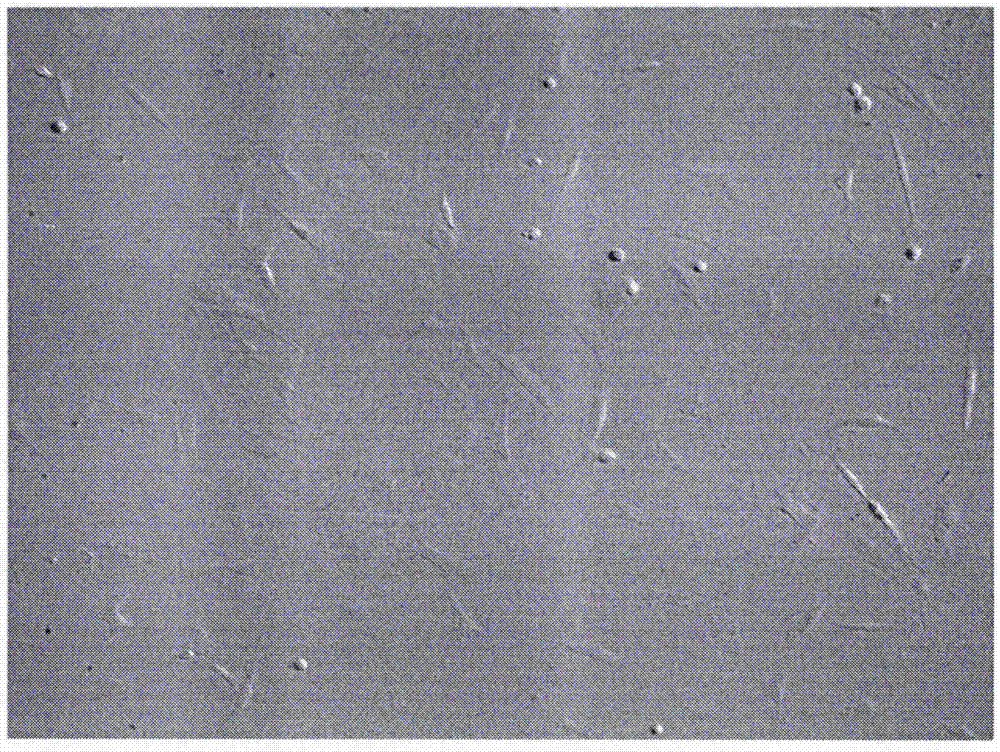
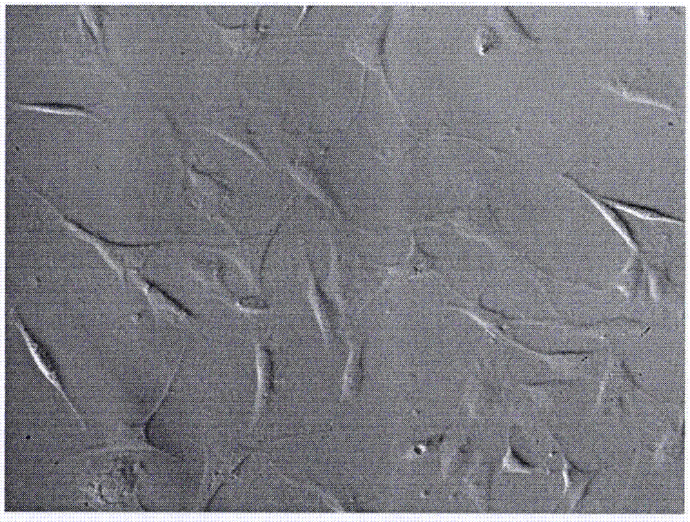
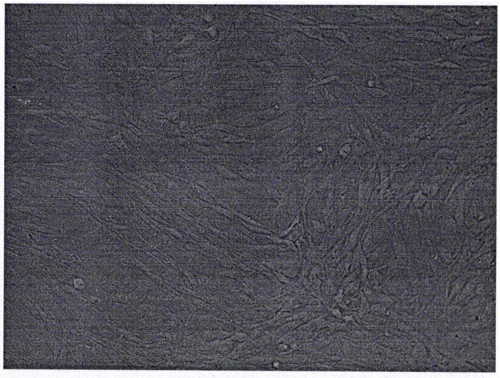
  • Method for separating and culturing mouse synovial cells
  • Method for separating and culturing mouse synovial cells
  • Method for separating and culturing mouse synovial cells

Examples


Embodiment Construction

[0037] The terms used in the present invention, unless otherwise specified, generally have the meanings commonly understood by those skilled in the art.

[0038] The present invention will be described in detail below in conjunction with the accompanying drawings and examples, and the protection content of the present invention is not limited to the following examples.

[0039] In the following examples, various procedures and methods not described in detail are conventional methods well known in the art.

[0040] The experimental instruments and reagents used in this experiment are as follows:

[0041] A set of surgical instruments, inverted microscope (XDS-1A, Shanghai), fluorescence microscope (Leica, USA), cryogenic centrifuge (TD24B-WS, Shanghai), ultra-low temperature refrigerator (Zhongke Meiling), pipette gun (Eppendorf, USA) ), electronic analytical balance (Sartorius, USA), ultra-clean bench (HJ-CJ-1D, Shanghai), CO 2 Cell culture incubator (SANYO MCO-17AI, Japan)....


PUM

PropertyMeasurementUnit
purityaaaaaaaaaa

Abstract

The invention provides a method for isolating and cultivating mouse cells, which includes the following steps: (1) sterilizing tissue processing equipment; (2) selecting male BALB / c mice aged 1 to 2 months, killing them by breaking their necks, and using surgical scissors. Separate the synovial layer and fibrous layer of the joint capsule, then remove the synovial layer tissue, put the tissue into pre-cooled sterile PBS solution containing double antibody and wash it several times to remove blood stains and fatty tissue; (3) Cut the tissue into pieces, and use Digest with preheated type II collagenase and trypsin respectively; (4) Stop digestion, centrifuge, and discard the supernatant; (5) Inoculate into the coated culture flask by manual adhesion method; (6) After incubation for 1 to 2 hours, Cultivate in a 37°C, 5% CO2 environment; (7) Cell morphology observation; (8) Cell subculture; (9) Cell identification. The method for isolating and culturing mouse synovial cells provided by the invention is simple to operate, has a large number of cells obtained, and has a high survival rate. It is an ideal primary method for isolating and cultivating mouse synovial cells and provides reliable results for experiments. Cell resources.

Description

Technical field [0001] The invention belongs to the field of cell culture technology of modern biotechnology, and specifically relates to a method for isolating and cultivating mouse synoviocytes. Background technique [0002] Synovium is the inner layer of the joint capsule. It is light red, thin and soft, and is composed of loose connective tissue. The synovial membrane secretes synovial fluid and plays an important role in joint movement. Normal synovium is divided into two layers, namely the thin cell layer (lumen layer) and the vascular layer (subintimal layer). [0003] Synovial cells originate from the mesenchyme in the blastema of bones. Synovial tissue contains a variety of cells, such as synoviocytes, leukocytes, adipocytes, etc. Synovial cells are divided into two types: type A and type B. Macrophage-like A-type cells (MLC) have filopodia and have phagocytic function but no proliferation ability. Type B synoviocytes (FLC), also known as fibroblast synoviocyte...

Claims


Application Information

Patent Timeline
Patent Type & Authority Applications(China)
IPC IPC(8): C12N5/071
CPCC12N5/0652C12N2501/33C12N2509/00C12N2533/32
Inventor 何刚张亚洲蒋敏齐来俊
Owner JIANGYIN CHI SCI
Learn More