
Fluorescent probe and its preparation method and use

A technology of fluorescent probes and detection reagents is applied in the field of preparation of fluorescent probes for distinguishing normal cells from cancer cells, and can solve the problems of difficult in vivo tumor imaging, inability to visualize in situ in real time, and low tissue penetration ability.

Active Publication Date: 2021-06-18
DALIAN UNIV OF TECH
View PDF0 Cites 0 Cited by
  • Summary
  • Abstract
  • Description
  • Claims
  • Application Information

AI Technical Summary

Problems solved by technology

[0004] Fluorescent probes have the advantages of high sensitivity, good selectivity, real-time in situ and visualized nondestructive testing, and can also overcome the shortcomings of traditional methods such as complicated sample pretreatment, expensive instruments, and inability to visualize in situ in real time. , fluorescent probes have been widely used in the field of molecular biological detection
However, so far, commercial kits for the detection of γ-glutaminyl transferase on the market use coumarin as a fluorescent group. This fluorescent probe needs to be excited by single photons, with short excitation wavelength and tissue penetration. Low power, difficult for in vivo tumor imaging applications
Especially based on targeting overexpressed γ-glutamine transferase, fluorescent probes to distinguish normal cells from mixed cultured cancer cells have not been reported in the literature

Method used


Image

  • Fluorescent probe and its preparation method and use
  • Fluorescent probe and its preparation method and use
  • Fluorescent probe and its preparation method and use

Examples


preparation example Construction

[0061] In another aspect, the present invention provides a method for preparing the above-mentioned fluorescent probe having the structure of formula I of the present invention, said method comprising the following steps:

[0062] (1) Compound 1 is reacted with malononitrile according to the molar ratio of 1:2 to 1:4 to prepare compound 2;

[0063]

[0064] (2) compound 3 and compound 4 were reacted according to the molar ratio of 1:1 to 1:2 to prepare compound 5;

[0065]

[0066] (3) Compound 5 is reacted with manganese dioxide according to a molar ratio of 1:5 to 1:10 to prepare compound 6;

[0067]

[0068] (4) compound 2 and compound 6 were reacted according to the molar ratio of 1:1 to 1:2 to prepare compound 7;

[0069]

[0070] (5) compound 7 reacts with trifluoroacetic acid according to the molar ratio of 1:50~1:200 to prepare the compound of formula I;

[0071]

[0072] In a specific embodiment, the above-mentioned fluorescent probe of the present in...

Embodiment 1

[0090] Solubility test of fluorescent probe in PBS (10mM pH 7.4) buffer solution.

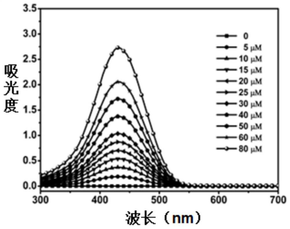
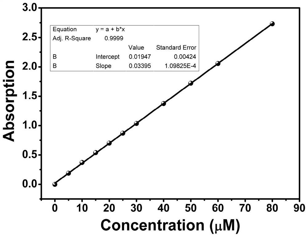
[0091] Prepare the mother solution of the fluorescent probe in a volumetric flask with dimethyl sulfoxide solvent. Then use a micro-sampler to take samples, make solutions with concentrations of 0, 5, 10, 15, 20, 25, 30, 40, 50, 60, and 80 μM, and scan them with an ultraviolet-visible absorption spectrometer to obtain the following: Figure 1a UV-Vis absorption spectrum. Take the absorption intensity (Abs) at the position of the maximum absorption peak as the ordinate, and the concentration as the abscissa to plot ( Figure 1b ). It was found that with the continuous increase of the concentration, the concentration and the absorption intensity showed a strong linear relationship (R 2 =0.9999), indicating that the fluorescent probe of the present invention has good solubility in PBS (10 mM pH 7.4) buffer solution.

Embodiment 2

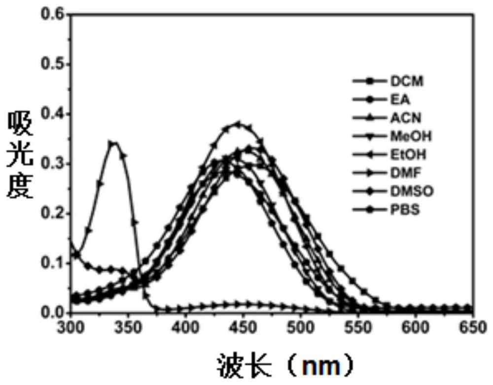
[0093] Investigate the UV-visible absorption and fluorescence emission experiments of fluorescent probes in different common solvents.

[0094] Add 5 μM fluorescent probes to dichloromethane (DCM), ethyl acetate (EA), acetonitrile (ACN), methanol (MeOH), ethanol (EtOH), dimethylsulfoxide (DMSO), dimethylformaldehyde Amide (DMF), PBS (10mM pH 7.4) buffer solution to scan its UV-Vis absorption spectrum ( Figure 2a ) and fluorescence spectra ( Figure 2b ). In most solvents, the position of the absorption peak of the probe is basically unchanged, only the absorption peak in DMF has an obvious blue shift. Fluorescent probes luminesce most strongly in DMSO solution.


PUM


Abstract

Fluorescent probe and its preparation method and use. The fluorescent probe has the structure of formula I. The fluorescent probe molecule of the above formula I structure of the present invention is a long-wavelength fluorescent probe with excellent two-photon performance designed based on the intramolecular charge transfer (Intramolecular Charge Transfer, ICT for short) mechanism. By targeting the overexpressed γ-glutamine transferase on the surface of cancer cells, the recognition site is selectively cleaved by the enzyme, thereby releasing a strong electron-donating group and enhancing the fluorescence at long wavelengths. This fluorescent probe has the advantages of good solubility, strong photostability, not affected by the acidity and alkalinity of the system, and two-photon excitation (excitation wavelength 800nm), and can realize selective confocal fluorescence imaging of cancer cells .

Description

technical field [0001] The invention belongs to the field of fine chemical industry, and relates to a preparation method and application of a fluorescent probe for distinguishing normal cells and cancer cells. Background technique [0002] With the deterioration of the living environment and the increase of life pressure, the number of human beings suffering from cancer diseases shows a trend of sharp increase. According to statistics, in 2015, there were 14.1 million new cancer patients and 8.2 million deaths worldwide. It is estimated that new cases will increase by 70% in the next 20 years. Due to untimely diagnosis and lack of effective personalized treatment plan, the treatment inefficiency rate of cancer patients is as high as 75%. For an ordinary family, it often brings a catastrophic blow. Although the government invests a large amount of research funding every year, it is expected to conquer the disease of treating cancer. But for now, the "early detection, early...

Claims


Application Information

Patent Timeline
Patent Type & Authority Patents(China)
IPC IPC(8): C07D307/68C09K11/06G01N21/64
CPCC07B2200/07C07D307/68C09K11/06C09K2211/1007C09K2211/1088G01N21/64
Inventor 彭孝军李海东姚起超樊江莉杜健军
Owner DALIAN UNIV OF TECH
Learn More