Glioma-targeting composite nanometer preparation, preparation method and applications thereof

A nano-formulation, glioma technology

Active Publication Date: 2018-04-10
NANJING MEDICAL UNIV
View PDF4 Cites 2 Cited by
  • Summary
  • Abstract
  • Description
  • Claims
  • Application Information

AI Technical Summary

Problems solved by technology

[0007] The purpose of the present invention is to address the defects of the existing nasal preparations for the treatment of glioma, and provide a nasal nanocomposite targeting glioma that delivers liver cancer-derived growth factor shRNA, its preparation method and its application

Method used


Image

  • Glioma-targeting composite nanometer preparation, preparation method and applications thereof
  • Glioma-targeting composite nanometer preparation, preparation method and applications thereof
  • Glioma-targeting composite nanometer preparation, preparation method and applications thereof

Examples


Embodiment 1

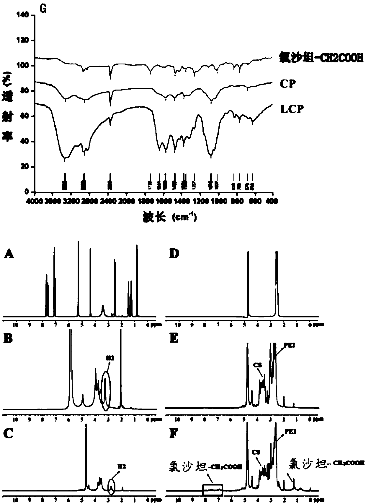
[0036] The synthesis of embodiment 1 carrier material LCP

[0037] ①Synthesis of CP copolymer: Dissolve chitosan and potassium periodate in pH 4.5 acetic acid / sodium acetate buffer respectively, mix the two solutions, and react with gentle stirring at 4°C under nitrogen protection. After 48 hours, add ethyl alcohol Diol (10%, v / v) continued to stir for 30 min. After the reaction was terminated, small molecular impurities were removed by dialysis bag method, and freeze-dried to obtain oxidized chitosan (O-CS), which was stored at -20°C.

[0038] ②Weigh an appropriate amount of O-CS and dissolve it in water, mix it with PEI (molecular weight 1.8k), and stir gently at 4°C under nitrogen protection. After reacting for 48 hours, add boron dissolved in sodium hydroxide into the reaction bottle three times. Sodium hydride, continue under nitrogen protection, stir at room temperature, take out the liquid after 12 hours of reaction, use dialysis (molecular weight cut-off 3.5kDa) to dia...

Embodiment 2

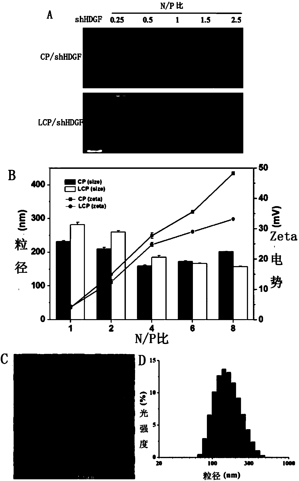
[0043] Example 2 Effect of different N / P on nanocomposite LCP / shHDGF experiment

[0044] Preparation method: Prepare LCP / shHDGF nanocomposites by mixing the copolymer LCP and shHDGF according to the N / P ratio of 0.25, 1, 2, 2.5, 4, 6, and 8. After mixing the two in equal volume, vortex for 1 min, to ensure Copolymer LCP and shHDGF can be fully compounded and used after standing at room temperature for 30 minutes. The preparation method of the control CP / shHDGF nanocomposite is the same as that of the LCP / shHDGF nanocomposite, except that LCP is replaced by CP.

[0045] Performance testing: The sample is placed in the sample cell, and the particle size, Zeta potential, and polydispersity coefficient are measured with a Malvern laser particle size analyzer Zetasizer Nano-S90. The morphology of the nanocomposites was observed and photographed by transmission electron microscopy.

[0046] The result is as figure 2 As shown in A: when N / P is 0.25, LCP and CP cannot completely c...

Embodiment 6

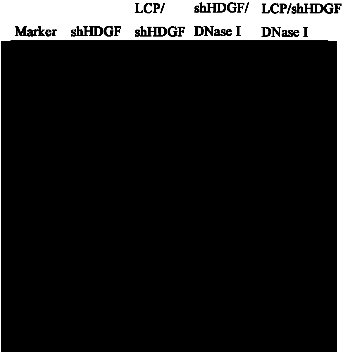
[0049] Anti-RNase Degradation Ability of Nasal Nanocomposite LCP / shHDGF and Release of shHDGF

[0050] Prepare the LCP / shHDGF nanocomplex with an N / P ratio of 8, then add 1 μL of RNase I, place the centrifuge tube containing the mixture in a constant temperature shaker, mix for 30 min at 37 °C and 100 rpm, and then add 4 μL Ethylenediaminetetraacetic acid (EDTA), after standing at 65°C for 20min, mix the sample with heparin (1000U / mL) at 37°C for 1h to release shHDGF from the complex, and finally use agarose gel Electrophoresis sample loading, 100V voltage for about 1h, use gel imaging system to observe and take pictures ( image 3 shown).

[0051] The results are as follows: After the LCP / shHDGF nanocomposite and naked shHDGF were incubated with RNase I for 30 min, the band of naked shHDGF disappeared, while the band of LCP / shHDGF nanocomposite still existed, indicating that the copolymer LCP has the ability to protect genes. In addition, as can be seen by competing with he...


PUM

PropertyMeasurementUnit
particle diameteraaaaaaaaaa
molecular weightaaaaaaaaaa

Abstract

The invention provides a glioma-targeting composite nanometer preparation, a preparation method and applications thereof, wherein the glioma-targeting composite nanometer preparation is a nasal nanometer complex assembled by coating a coated material hepatoma-derived growth factor delivery shRNA with a coating material losartan-chitosan-polyethyleneimine vector copolymer, and the particle size is50-250 nm. The preparation method comprises: carrying out an oxidation reaction on chitosan and potassium periodate to obtain oxidized chitosan; carrying out a reaction on the oxidized chitosan and polyethyleneimine to obtain a chitosan-polyethyleneimine copolymer; carrying out a Schiff base reaction on the chitosan-polyethyleneimine copolymer and carboxylated losartan to obtain a losartan-chitosan-polyethyleneimine vector copolymer; and assembling the losartan-chitosan-polyethyleneimine vector copolymer and shHDGF to obtain the nasal nanometer complex. According to the present invention, after the nasal cavity product is prepared from the glioma-targeting composite nanometer preparation, the good brain glioma targeting property can be provided.

Description

technical field [0001] The present invention relates to the field of pharmaceutical preparations, in particular to a glioma-targeting nasal nanocomposite delivering liver cancer-derived growth factor shRNA, a preparation method and use thereof. Background technique [0002] Malignant glioma is the most common primary tumor of the central nervous system, accounting for about 80% of primary malignant brain tumors, with extremely high recurrence and mortality rates. The rapid and invasive growth throughout the whole brain is a prominent biological feature of malignant glioma, and it is also the biggest obstacle to its treatment and the root cause of poor prognosis. Clinically, even if surgical treatment is combined with radiotherapy, chemotherapy, etc. The average survival period is only about 15 months. There is still no effective treatment for malignant glioma. Therefore, it is of great significance to conduct research on the treatment of glioma. [0003] Gene therapy for ...

Claims


Application Information

Patent Timeline
Patent Type & Authority Applications(China)
IPC IPC(8): A61K47/61A61K47/59A61K31/713A61K31/4178A61K38/18A61P35/00
CPCA61K9/0043A61K31/4178A61K31/713A61K38/18A61K2300/00
Inventor 李瑞张爱霞张明晚周美玲苏平黄元沈鸿
Owner NANJING MEDICAL UNIV
Why Eureka
Try Eureka