Mechanical stimulation method for improving in-vitro preservation effect of articular cartilage
A technology of articular cartilage and mechanics, applied in the field of biomedicine, can solve the problems of decreased activity of chondrocytes, achieve the effects of reducing chondrocyte apoptosis, improving utilization rate and cartilage repair technology level, and improving preservation effect
- Summary
- Abstract
- Description
- Claims
- Application Information
AI Technical Summary
Problems solved by technology
Method used
Image

Examples
preparation example Construction
[0039] The tissue culture medium used in the present invention can also be TSMU culture medium, and the TSMU culture medium is a published Chinese patent application "a preservation solution for osteochondral grafts and its preparation method" (application number: CN201510093497.0) Osteochondral graft preservation solution described in , each 1000ml of osteochondral graft preservation solution includes: glucose 80-125mmol, amino acid 0.1-2.0mmol, antioxidant 0.2-2.0mmol, basic fibroblast growth factor 5-60nmol , Inorganic salt 1-120mmol, vitamin 1-9mmol, sodium pyruvate 1-2mmol, penicillin 50-70U, the rest is deionized water. The inorganic salt is selected from one or a combination of sodium chloride, potassium chloride and magnesium chloride. The specific preparation steps of the osteochondral graft preservation solution used in the present invention are:
[0040] Take glucose 14.4g, amino acid 0.21g, antioxidant 0.23g, basic fibroblast growth factor 0.0016g, sodium chloride...
Embodiment 1
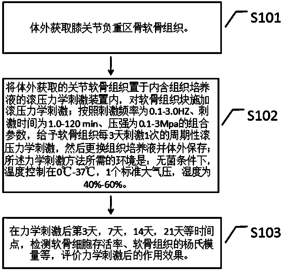
[0048] (1) In vitro aseptically obtain fresh osteochondral tissue in the weight-bearing area of the knee joint within 3 hours of animal death, with a length of 15 mm and a diameter of about 6.0 mm.
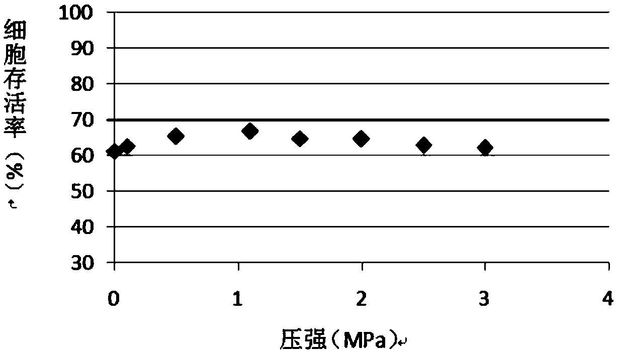
[0049] (2) Mechanical stimulation scheme at the initial stage of the experiment: place the articular cartilage tissue obtained in vitro in a rolling mechanical stimulation device containing DMEM tissue culture medium, and apply rolling mechanical stimulation to the cartilage tissue block; according to the stimulation frequency of 2HZ, the stimulation time The combined parameters of 30min and pressure of 1.5Mpa give cartilage mechanical stimulation, and then replace with fresh tissue culture medium and store in vitro; the environment required for the mechanical stimulation method is: under sterile conditions, the temperature is controlled at 0°C-37 ℃, 1 standard atmospheric pressure, humidity 40%-60%.
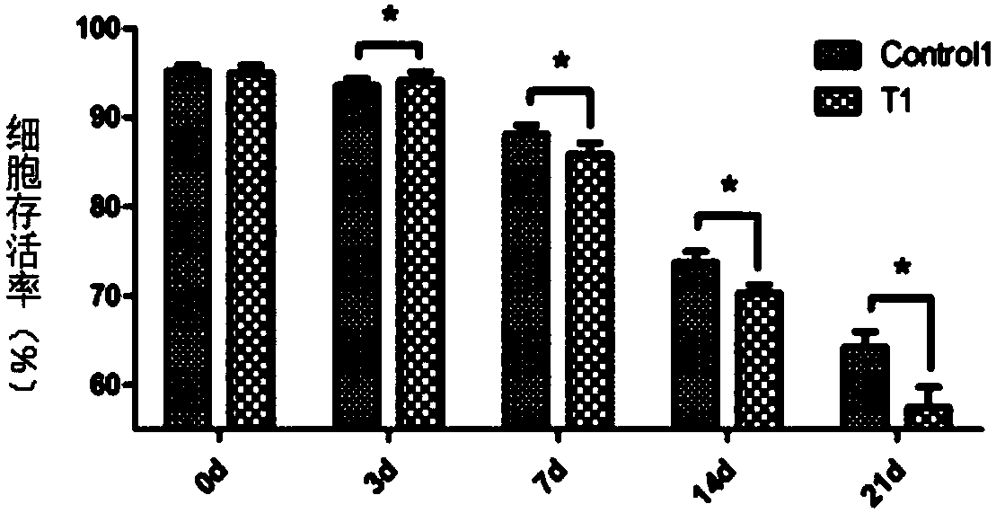
[0050] (3) On the 3rd day, 7th day, 14th day, and 21st day after mechanical s...
Embodiment 2
[0052] (1) Fix the knee joint sample on the operating table in accordance with the aseptic principle, disinfect the sample with povidone iodine, and spread a sterile towel. Make a midline incision of the knee joint, open the joint capsule, dissect the patellar ligament and the anterior and posterior cruciate ligaments, select the load-bearing area of the medial and medial condyle of the femoral articular surface, and use a special osteochondral harvesting instrument to collect cylindrical osteochondral pieces (about 10 mm in length and 8 mm in diameter) , given continuous PBS washing during the sampling process), and then the bottom surface of the osteochondral block was trimmed and leveled, put into a sterile vessel and rinsed with PBS.
[0053] (2) Using the developed rolling pressure loading device, the cartilage tissue is loaded according to the loading frequency, time, and pressure as the loading conditions; the loading box is sterilized with ethylene oxide; the conditio...
PUM
| Property | Measurement | Unit |
|---|---|---|
| Length | aaaaa | aaaaa |
| Diameter | aaaaa | aaaaa |
| Diameter | aaaaa | aaaaa |
Abstract
Description
Claims
Application Information
 - R&D
- Intellectual Property
- Life Sciences
- Materials
- Tech Scout
- Unparalleled Data Quality
- Higher Quality Content
- 60% Fewer Hallucinations
Browse by: Latest US Patents, China's latest patents, Technical Efficacy Thesaurus, Application Domain, Technology Topic, Popular Technical Reports.
© 2025 PatSnap. All rights reserved.Legal|Privacy policy|Modern Slavery Act Transparency Statement|Sitemap|About US| Contact US: help@patsnap.com