Capillary micro-droplet metal ball detection method for surface enhanced Raman spectrum
A technology of surface-enhanced Raman and detection methods, applied in the field of sensitive detection and analysis, can solve the problems of large volume and difficult laser focusing, and achieve the effects of simple operation, real-time quantitative detection, and less spectral spurious peaks.
- Summary
- Abstract
- Description
- Claims
- Application Information
AI Technical Summary
Problems solved by technology
Method used
Image

Examples
Embodiment 1
[0051] A method for detecting capillary micro-droplet metal balls for surface-enhanced Raman spectroscopy, the steps are as follows:
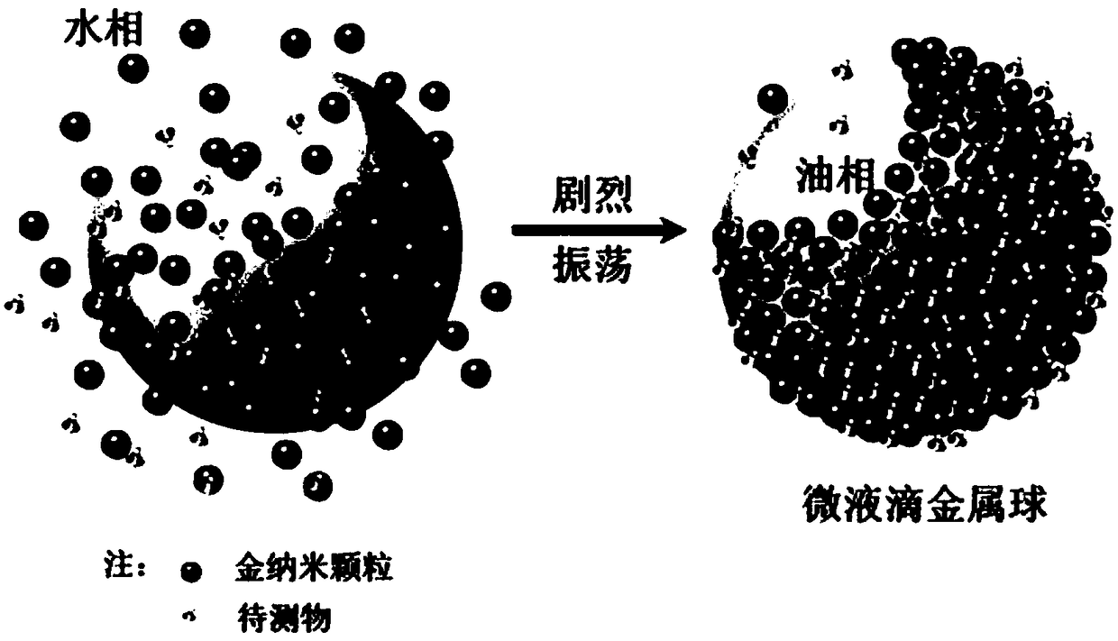
[0052] (1) Mix the noble metal nano-sol with an organic solvent with a density greater than water, and then add the extract of the analyte to shake vigorously;
[0053] (2) The noble metal nanomaterials in the noble metal nanosol are quickly assembled at the oil-water interface to form micro-droplet metal balls with adjustable gaps between nanomaterials;
[0054] Such as figure 1 The enlarged schematic diagram is shown, gold nanoparticles and analyte molecules are quickly assembled at the oil-water interface to form micro-droplet metal balls with adjustable gaps between nanomaterials;
[0055] (3) Utilize capillary action to suck the micro-droplet metal ball into the capillary;
[0056] (4) Place the above-mentioned capillary adsorbed with micro-droplet metal balls under a Raman spectrometer for detection, and the SERS characteristic fingerpr...
Embodiment 2
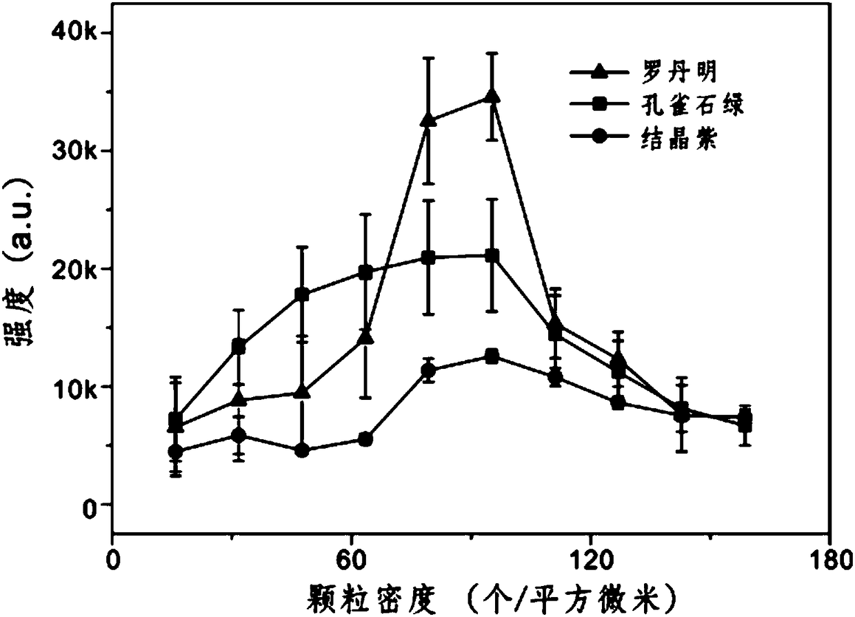
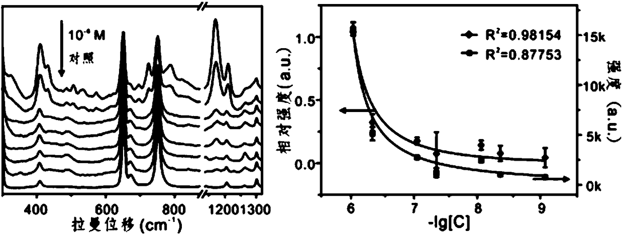
[0067] Add 30μL o-dichloroethane containing malachite green and 0.9mL gold particles with a diameter of 80nm (the enhancement effect of SERS is the best at this time) into the container vial treated with hydrophilicity, and shake vigorously to assemble into micro-droplet metal balls . Then transferred to the capillary for Raman detection, the concentration of malachite green was 1×10 -6 M, 5×10 -7 M, 1×10 -7 M, 5×10 -8 M, 1×10 -8 M, 5×10 -9 M , 1×10 -9 M and 0 M, such as image 3 shown. The Raman characteristic peaks of malachite green are: 436 cm -1 , 788cm -1 、896 cm -1 、1174 cm -1 、1367 cm -1 and 1616 cm -1 . Choose 1174 cm -1 The Raman peak at 656 cm -1 The o-dichloroethane peak at the place is used as the internal standard, and the result shows that when no internal standard is added, R 2 = 0.87753, R after internal standard treatment 2 = 0.98154. Raman parameters include: microscope objective lens × 20, excitation wavelength 785 nm, detection...
Embodiment 3
[0069] Crucian carp meat was crushed and homogenized, and then malachite green (MG) dissolved in o-dichloroethane was added to make malachite green concentrations of 0 M, 2.0×10 -8 M , 5.0×10 -8 M , 1.0×10 -7 M and 2.0×10 -7 M samples. Finally, an extract containing malachite green was prepared, and the preparation process was as follows: (1) Take 4.00±0.04g sample and mix with 1000μL of 9.5g / L hydroxylamine solution to prevent MG degradation, and react at room temperature for 15 minutes before extraction; (2) Add 2.0±0.2g anhydrous magnesium sulfate to the homogenate and vortex vigorously for 1 minute; (3) Add 4.0±0.1g aluminum oxide to the homogenate and vortex vigorously for 30 seconds to remove lipids in the sample; (4) Take the supernatant and put it in a centrifuge tube, and centrifuge at 15000rpm for 10 minutes. After centrifugation, transfer the supernatant to a 20mL test tube dried under nitrogen at 50°C, add 2.0±0.1g alumina, vortex for 30 seconds, then trans...
PUM
| Property | Measurement | Unit |
|---|---|---|
| diameter | aaaaa | aaaaa |
Abstract
Description
Claims
Application Information
 - R&D
- Intellectual Property
- Life Sciences
- Materials
- Tech Scout
- Unparalleled Data Quality
- Higher Quality Content
- 60% Fewer Hallucinations
Browse by: Latest US Patents, China's latest patents, Technical Efficacy Thesaurus, Application Domain, Technology Topic, Popular Technical Reports.
© 2025 PatSnap. All rights reserved.Legal|Privacy policy|Modern Slavery Act Transparency Statement|Sitemap|About US| Contact US: help@patsnap.com