Preparation method of ovalbumin nano particle loaded with EGCG (Epigallocatechin Gallate), and application thereof
A technology of ovalbumin and nanoparticles, which is applied in the field of preparation of EGCG-loaded ovalbumin nanoparticles, which can solve the problems of poor stability, easy oxidation, and decomposition, and achieve the effects of reducing oxidative decomposition, easy cell phagocytosis, and increasing half-life
- Summary
- Abstract
- Description
- Claims
- Application Information
AI Technical Summary
Problems solved by technology
Method used
Image

Examples
Embodiment 1
[0033] Embodiment 1 is the preparation method of loading EGCG ovalbumin nanoparticles, comprising the following steps:
[0034] (1) Weigh ovalbumin (OVA) into a 50 mL centrifuge tube, then add PBS buffer solution with pH 6.6, vortex to fully dissolve, and hydrate overnight;
[0035] (2) Centrifuge the ovalbumin solution hydrated overnight and collect the supernatant to remove insoluble impurities;
[0036] (3) Keep the solution obtained in step (2) in a water bath at 80°C for 20 min;
[0037] (4) After the solution in step (3) is bathed in water, add EGCG to the solution immediately, and the solution becomes an emulsion at this time;
[0038] (5) Vortex the EGCG-containing emulsion obtained in step (4) for 30 s, then immediately cool to room temperature with running water;
[0039] (6) Immediately put the emulsion obtained in step (5) into an ice bath, and sonicate the probe for 1 min;
[0040] (7) Centrifuge the emulsion obtained in step (6) at a low speed and collect the ...
Embodiment 2
[0044] Experiment 1: The morphology of nanoparticles was observed by field emission scanning electron microscopy. First, suspend the lyophilized nanoparticles in secondary water or PBS buffer, and dilute to an appropriate concentration, then drop the suspension onto a silicon wafer and air-dry it. Before analysis, the nanoparticles need to be vacuum-sprayed with platinum.
[0045] figure 1 Field emission scanning electron microscope image of nanoparticles. It can be seen from the obtained electron microscope photos that the nanoparticles are spherical, the particle size distribution is relatively uniform, and they are densely packed, which is also the characteristic of protein nanoparticles.
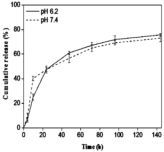
[0046] Test 2: In order to simulate the in vivo release of the drug, the release solution uses phosphate buffer solution with pH = 7.4 and pH = 6.8 to simulate the pH in the body. First, accurately weigh an appropriate amount of nanoparticles (the mass of the nanoparticles is calculate...
PUM
 Abstract
Description
Claims
Application Information
 - R&D
- Intellectual Property
- Life Sciences
- Materials
- Tech Scout
- Unparalleled Data Quality
- Higher Quality Content
- 60% Fewer Hallucinations
Browse by: Latest US Patents, China's latest patents, Technical Efficacy Thesaurus, Application Domain, Technology Topic, Popular Technical Reports.
© 2025 PatSnap. All rights reserved.Legal|Privacy policy|Modern Slavery Act Transparency Statement|Sitemap|About US| Contact US: help@patsnap.com