
Preparation method of biogenic small-aperture tissue engineering blood vessels

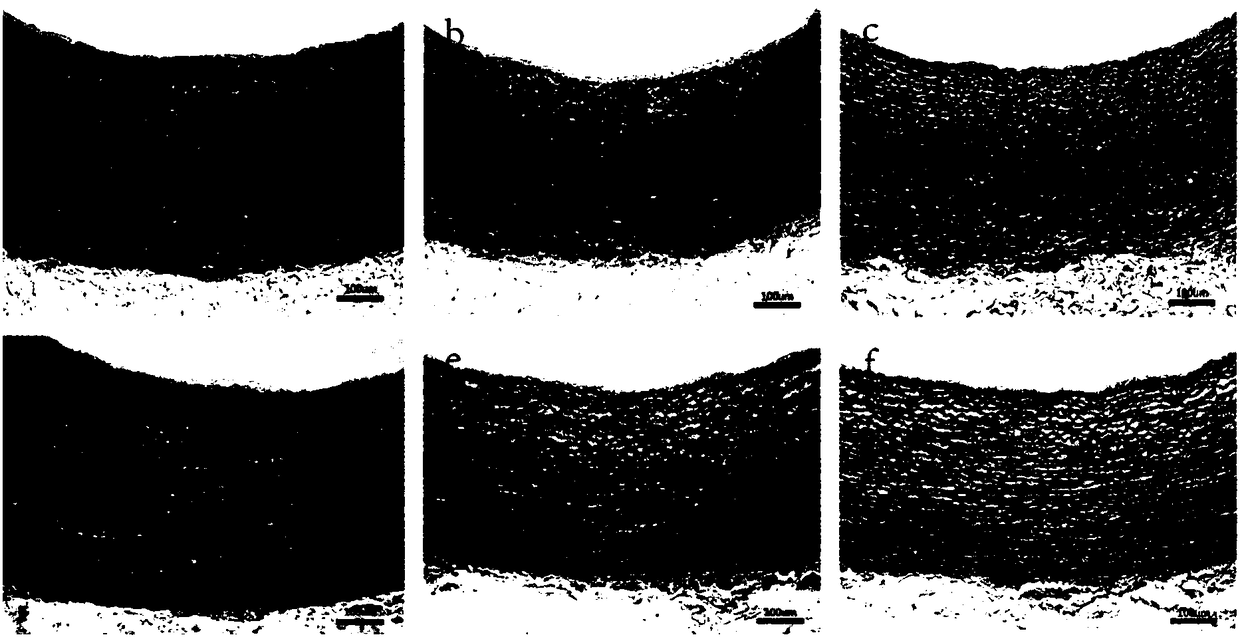
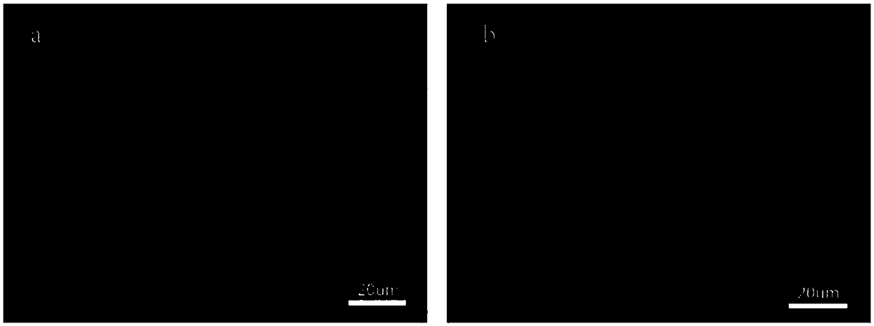
A tissue engineering and biologically derived technology, applied in tissue regeneration, medical science, prosthesis, etc., can solve the problems of occlusion, hyperplasia of organ cavity, destruction of biological characteristics of vascular matrix, etc., to avoid the residual of toxic substances and strengthen the decellularization effect. , to avoid the effect of degradation

Active Publication Date: 2018-06-12
XUANWU HOSPITAL OF CAPITAL UNIV OF MEDICAL SCI
View PDF13 Cites 7 Cited by
  • Summary
  • Abstract
  • Description
  • Claims
  • Application Information

AI Technical Summary

Problems solved by technology

[0009] Although the mechanical strength of the decellularized matrix can be improved by cross-linking, the biological properties of the cross-linked vascular matrix are damaged, which is not conducive to the adhesion, invasion and growth of cells in vivo. Affect the compliance of the vascular matrix, easily cause intimal hyperplasia and occlusion of the lumen at the suture site at both ends of the stent, and affect the long-term survival of the acellular matrix in the body
In addition, the cross-linked vascular matrix is ​​more prone to immune reactions, resulting in severe fibrosis

Method used


Image

  • Preparation method of biogenic small-aperture tissue engineering blood vessels
  • Preparation method of biogenic small-aperture tissue engineering blood vessels
  • Preparation method of biogenic small-aperture tissue engineering blood vessels

Examples


Embodiment 1

[0053] Example 1: Preparation of tissue engineered blood vessels

[0054] 1. Materials

[0055] Fresh animal blood vessels: fresh porcine carotid artery vessels (porcine iliac artery vessels can also be used)

[0056] 2. Method

[0057] (1) freeze-thaw

[0058] Place the fresh animal blood vessels in PBS buffer containing 0.1% EDTA, place them in a -80°C deep-low temperature refrigerator for 2 hours, and place them in a 37°C water bath for 20 minutes immediately after taking them out. Repeat the above operation twice, that is, perform a total of 3 freeze-thaw cycles;

[0059] (2) Remove the cell debris that fell off after freezing and thawing

[0060] The blood vessel material obtained in step (1) is placed in distilled water and fully rinsed at 4°C to remove detached cell residues;

[0061] (3) Detergent decellularization

[0062] The vascular material obtained in step (2) was soaked in Tris-HCl buffer containing 0.1% EDTA and 1% TritonX-100 and shaken at room temperatu...

Embodiment 2

[0096] Example 2: Preparation of tissue engineered blood vessels

[0097] 1. Materials

[0098] Fresh animal blood vessels: porcine carotid arteries or porcine iliac arteries

[0099] 2. Method

[0100] (1) freeze-thaw

[0101] Place fresh animal blood vessels in PBS buffer containing 0.02% EDTA, place them in a deep freezer at -80°C for 1 hour, and place them in a 37°C water bath for 10 minutes immediately after taking them out. Repeat the above operation once, that is, perform a total of 2 freeze-thaw cycles;

[0102] (2) Remove the cell debris that fell off after freezing and thawing

[0103] The blood vessel material obtained in step (1) is placed in distilled water and fully rinsed at 4°C to remove detached cell residues;

[0104] (3) Detergent decellularization

[0105] The vascular material obtained in step (2) was soaked in Tris-Hcl buffer containing 0.02% EDTA and 1% TritonX-100 and shaken at room temperature for 48 hours, and then fully washed with distilled wa...

Embodiment 3

[0108] Example 3: Preparation of tissue engineered blood vessels

[0109] 1. Materials

[0110] Fresh animal blood vessels: porcine iliac arteries or porcine carotid arteries

[0111] 2. Method

[0112] (1) freeze-thaw

[0113] Place fresh animal blood vessels in PBS buffer containing 0.2% EDTA, place them in a -80°C deep-low temperature refrigerator for 3 hours, and place them in a 37°C water bath for 30 minutes immediately after taking them out. Repeat the above operation 4 times, that is, a total of 5 freeze-thaw cycles;

[0114] (2) Remove the cell debris that fell off after freezing and thawing

[0115] The blood vessel material obtained in step (1) is placed in distilled water and fully rinsed at 4°C to remove detached cell residues;

[0116] (3) Detergent decellularization

[0117] The vascular material obtained in step (2) was soaked in Tris-Hcl buffer containing 0.2% EDTA and 1% TritonX-100 and shaken at room temperature for 12 hours, and then fully washed with ...


PUM


Abstract

The invention relates to a preparation method of biogenic small-aperture tissue engineering blood vessels, and belongs to the fields of tissue engineering and biological materials. The preparation method of the biogenic small-aperture tissue engineering blood vessel comprises the following steps: (1) freeze thawing: placing fresh animal blood vessels into a PBS (Phosphate Buffer Solution) containing 0.02 to 0.2 percent of EDTA (Ethylene Diamine Tetraacetic Acid), and performing quick freezing at the temperature of 80 DEG C below zero for 1 to 3 hours, taking the fresh animal blood vessels out,and immediately performing unfreezing at the temperature of 37 DEG C for 10 to 30 minutes; repeating the abovementioned operations for 2 to 5 times to obtain a blood vessel material; (2) removing cell debris fallen after the freeze thawing; (3) removing cells; (4) removing a detergent. The invention has the advantage that a cell removing method and a reagent composition which can effectively remove cell components in blood vessel tissues and protect the structural integrity of acellular matrixes to the maximum extent are provided.

Description

technical field [0001] The invention relates to a preparation method of biologically derived small-diameter tissue engineering blood vessels, belonging to the field of tissue engineering and biological materials. Background technique [0002] Cardiovascular disease has become the primary cause of threats to human health. In the treatment of arteriosclerosis, bypass grafting has always been the most effective treatment. For a long time, autologous blood vessels have been used as the gold standard for the treatment of small-caliber vascular diseases. 1 / 3 of patients cannot find a suitable autologous blood vessel due to vascular disease or previous vascular surgery. In the treatment of small-diameter vascular diseases, such as the treatment of coronary arteries and sub-knee vessels, artificial blood vessels are likely to cause blockage in the graft cavity due to inactivity, susceptibility to infection, poor compliance, and lack of regenerative capacity, and the long-term patenc...

Claims


Application Information

Patent Timeline
IPC IPC(8): A61L27/36A61L27/50
CPCA61L27/3633A61L27/3687A61L27/3691A61L27/507A61L2430/40
Inventor 谷涌泉王聪成津
Owner XUANWU HOSPITAL OF CAPITAL UNIV OF MEDICAL SCI
Learn More