Large visual field non-diffracting Bessel light sheet microscopic scanning imaging method and system

A Bessel light sheet and scanning imaging technology, which is applied in fluorescence/phosphorescence, material analysis through optical means, material excitation analysis, etc., can solve problems such as side lobe effects, achieve low photobleaching, and improve imaging quality.

Inactive Publication Date: 2018-07-20
HUAZHONG UNIV OF SCI & TECH RES INST SHENZHEN +1
View PDF7 Cites 10 Cited by
  • Summary
  • Abstract
  • Description
  • Claims
  • Application Information

AI Technical Summary

Problems solved by technology

However, when using a Bessel light sheet for illumination, although the Bessel beam itself has the characteristics of high central intensity, self-recovery effect, and anti-scattering, its cross-section is annular and has serious side lobe effects. In order to maintain a large field of view and improve axial resolution under aperture detection, effective suppression of the side lobes of Bessel light will become an inevitable focus and difficulty

Method used


Image

  • Large visual field non-diffracting Bessel light sheet microscopic scanning imaging method and system
  • Large visual field non-diffracting Bessel light sheet microscopic scanning imaging method and system
  • Large visual field non-diffracting Bessel light sheet microscopic scanning imaging method and system

Examples


Embodiment 1

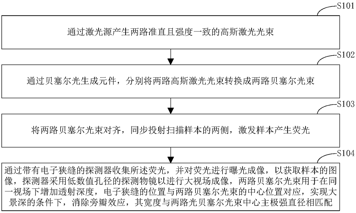
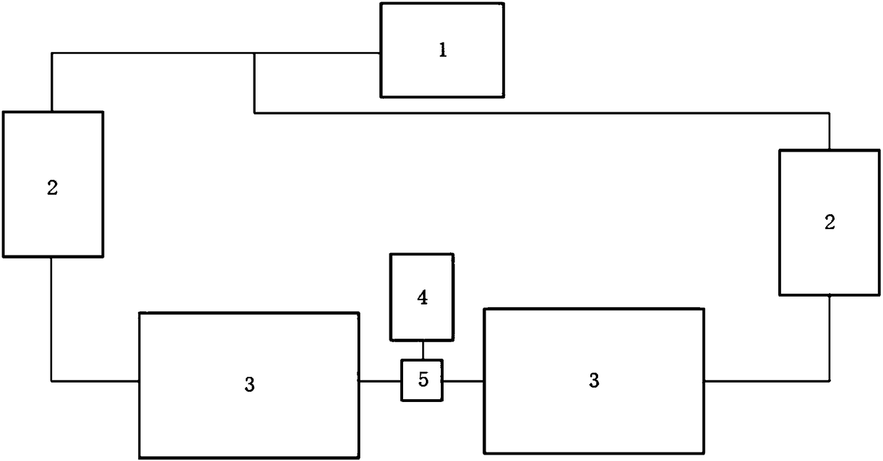
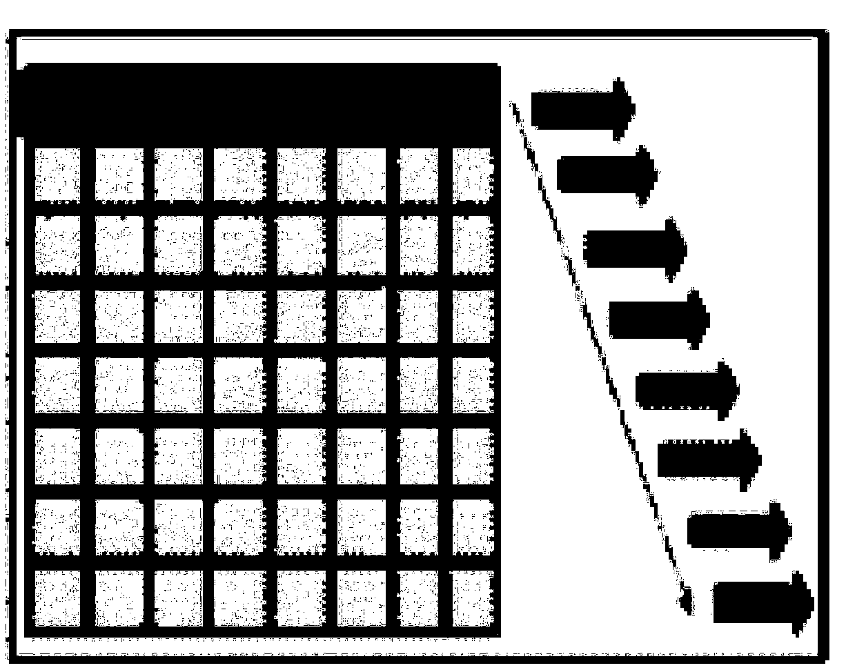
[0061] The embodiment of the present invention provides a large field of view non-diffraction Bessel light sheet micro-scanning imaging method, which strictly divides a laser light source into two beams of scanning lasers, and generates images under a large field of view through the Bessel light sheet generation mechanism. The Bezier light sheet, which implements a strictly synchronous scanning method. For transparent samples: the formed ultra-thin light sheet passes through the sample, excites the fluorescence, and simultaneously exposes and outputs an image through the image sensor.

[0062] In this embodiment, by dividing a stable light source into two paths and then combining them with attenuation plates, it is strictly guaranteed that the properties of the two paths of laser light are the same, including: frequency, light intensity, etc.

[0063] In this embodiment, the structure is generated by the light sheet, and the input Gaussian laser is converted into a high-intens...

Embodiment 2

[0075] Transgenic GFP-labeled mice were used as observation objects, and fluorescent signals were expressed in nerve cells, such as cortical pyramidal neurons, astrocytes, and deeper hippocampal cells. Nerve cells emit green fluorescence at an excitation wavelength of 488 nm. If you use an ordinary wide-field illumination inverted fluorescence microscope to observe, due to the extensive out-of-focus light irradiation, the image contrast is extremely low and the light pollution is serious; if you use an ordinary light-sheet microscopic imaging device, you must use an extremely thick light to cover the entire field of view of the mouse brain. If the optical film is thinner, the axial resolution will be greatly reduced. If a thinner optical film is used to achieve a higher axial resolution, the field of view that can be covered will be greatly reduced. However, the large-field non-diffraction Bessel light sheet microscopic scanning imaging method is adopted, which combines the no...


PUM


Abstract

The invention discloses a large visual field non-diffracting Bessel light sheet microscopic scanning imaging method and system. The method comprises following steps: generating two collimated Gauss laser beams with same intensity by a laser source; converting two Gauss laser beams into two Bessel beams by a Bessel light generating element respectively; aligning two Bessel beams, projecting the Bessel beams on two sides of a scanned sample to excite the sample to generate fluorescence; collecting the fluorescence by a detector with an electron slit, using the fluorescence to carry out exposureimaging to obtain the image of the sample, and adopting a detection object lens with a low value aperture to carry out large visual field imaging by the detector. Two Bessel beams are used to enhancethe transmission depth in a same visual field, the position of the electron slit is opposite to the center of two Bessel beams, under a condition of large depth of field, the side lobe effect is eliminated, and the depth of the electron slit is matched with the diameter of the center of two Bessel beams. The provided method and system can realize large visual filed and large depth of field imagingand improves the imaging quality of sample images.

Description

technical field [0001] The invention belongs to the technical field of microscopic imaging, and more particularly relates to a large field of view non-diffraction Bessel light sheet microscopic scanning imaging method and system. Background technique [0002] Light-sheet illumination microscopic technology, which uses a light beam sheet to excite fluorescence from the side of the sample, and obtains fluorescence images at the illumination level through a microscope objective lens and a CCD or sCMOS camera in the direction perpendicular to the light sheet, is an emerging technology in the 21st century. The cutting-edge imaging method, compared with the traditional epi-fluorescence microscope, has the characteristics of low phototoxicity, high temporal and spatial resolution, and high throughput, which makes it possible to observe the three-dimensional and dynamic panorama of organisms. At present, light-sheet microscopy has been regarded as a new imaging method, and has attra...

Claims


Application Information

Patent Timeline
Patent Type & Authority Applications(China)
IPC IPC(8): G01N21/64
CPCG01N21/6402
Inventor 费鹏王雪纯方春钰初婷婷赵方朱兰馨
Owner HUAZHONG UNIV OF SCI & TECH RES INST SHENZHEN
Why Eureka
Try Eureka