A quantum dot-magnetic bead miRNA sensor based on dsn enzyme and its preparation method and detection method

A quantum dot and sensor technology, applied in the field of nano-biomedicine, can solve the problem that single-stranded DNA and RNA have no effect, and achieve the effect of wide linear range, high operation sensitivity, and high detection sensitivity

Active Publication Date: 2021-05-18
NANJING UNIV OF POSTS & TELECOMM
View PDF10 Cites 0 Cited by
  • Summary
  • Abstract
  • Description
  • Claims
  • Application Information

AI Technical Summary

Problems solved by technology

[0005] Double-strand-specific nuclease (Duplex-specific nuclease or DSN) can highly selectively recognize and cut DNA in double-stranded DNA or DNA-RNA double-strand, but has little effect on single-stranded DNA and RNA

Method used


Image

  • A quantum dot-magnetic bead miRNA sensor based on dsn enzyme and its preparation method and detection method
  • A quantum dot-magnetic bead miRNA sensor based on dsn enzyme and its preparation method and detection method
  • A quantum dot-magnetic bead miRNA sensor based on dsn enzyme and its preparation method and detection method

Examples


Embodiment 1

[0048] Add 22.5 μL of SA-QD (Streptavidin QD) with a concentration of 1 μM, 22.5 μL of Biotin-DNA with a concentration of 10 μM, 200 μL of blocking solution, and 600 μL of Tris-HCl buffer solution into a 1.5 mL low adsorption centrifuge tube and shake at a speed of 6000 rpm / min for 1 hour , construct the QD-DNA probe, and then ultrafilter at 4000rpm for 5min to remove the DNA not connected with QD, and repeat the ultrafiltration process 3 times. After the last ultrafiltration until 100 μL of the QD-DNA probe solution remained, 75 μL of Tris-HCl buffer and 50 μL of blocking solution were added to obtain a high-concentration QD-DNA probe. Divide the constructed high-concentration QD-DNA probe into 10 equal parts: take 22.5 μL QD-DNA probe for each part, add 135 μL Tris-HCl buffer solution and 67.5 μL blocking solution.

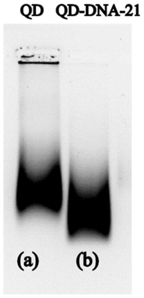
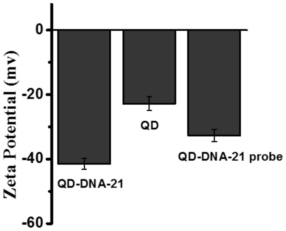
[0049] The surface of quantum dots is negatively charged, and the surface of DNA is negatively charged. After QD is connected to DNA, the negative charge densit...

Embodiment 2

[0051] Before the experiment, wash the MB with ligation-washing buffer (B&W Buffer) 3 to 4 times to remove substances such as sodium azide in the mother liquor, take 45 μL of MB with a concentration of 10 mg / mL, 45 μL of Biotin-DNA with a concentration of 10 μM, and 600 μL Tris-HCl buffer solution was added to a low-adsorption centrifuge tube and shaken at a speed of 6000 rpm / min for 2 hours to construct the MB-DNA probe. Because the particle size of MB is relatively large (particle size is 1 μm), sedimentation is easy to occur, so the solution in the centrifuge tube was mixed every 30 minutes, so that MB and DNA can be fully connected. The constructed MB-DNA probe was magnetically separated and washed three times, and 225 μL of Tris-HCl buffer was added to obtain a high-concentration MB-DNA probe. Divide 10 equal parts of the MB-DNA probe solution: take 22.5 μL of high-concentration MB-DNA probe solution for each part, and then add 227.5 μL of hybridization buffer to make the...

Embodiment 3

[0054] Take 12 μL of 1 μM SA-QD (Streptavidin QD), 30 μL of Biotin-DNA with a concentration of 10 mM, 200 μL of blocking solution, and 600 μL of Tris-HCl buffer, and add them to a 1.5 mL low-adsorption centrifuge tube and shake at 6000 rpm / min for 2 hours , to construct QD-DNA bioprobes. Transfer the ligation product to an ultrafiltration tube and ultrafilter at a speed of 4000rpm for 5 minutes to remove as much as possible the DNA that is not connected to the QD in the supernatant. Ultrafilter three times in a row, take out the ultrafiltration tube and mix the supernatant with a pipette Prevent QDs from sticking to the filter membrane. Take out the supernatant and dilute the solution to 550 μL with ultrapure water, and prepare a QD solution of the same concentration with ultrapure water.

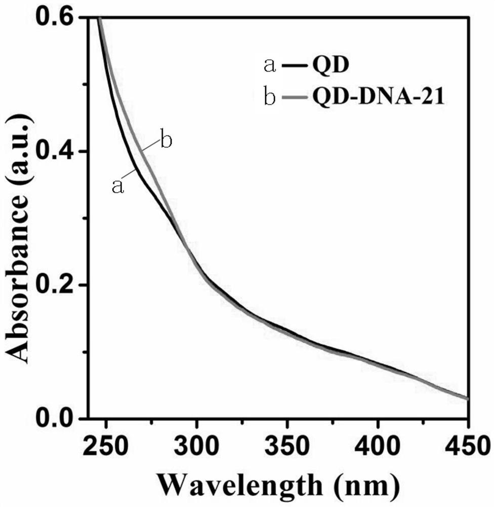
[0055] DNA and QD are coupled together by streptavidin and biotin, and the characteristic absorption peak of DNA can be observed by UV characterization of QD-DNA probe. From figure 2 It...


PUM


Abstract

The invention discloses a quantum dot-magnetic bead miRNA sensor based on DSN enzyme and its preparation method and detection method, through the covalent coupling of streptavidin on the surface of quantum dots and magnetic beads and biotin modified at the end of DNA Connect to construct QD‑DNA bioprobe and MB‑DNA bioprobe. The present invention utilizes the excellent optical properties of quantum dots, and the magnetic separation based on magnetic beads is more time-saving and more ideal in separation effect, and utilizes DSN enzymes to recognize and cut perfectly matched DNA double strands or RNA / DNA hybrids with high selectivity The DNA in the double strand can realize the characteristics of signal amplification, and a quantum dot-magnetic bead miRNA sensor based on DSN enzyme is prepared. The invention can realize highly sensitive and specific detection of miRNA, and will have potential applications in the fields of molecular biology, medicine and the like.

Description

technical field [0001] The invention belongs to the field of nano-biomedicine, and in particular relates to a DSN enzyme-based quantum dot-magnetic bead miRNA sensor and a preparation method and a detection method thereof. Background technique [0002] Micro ribonucleic acid (microRNA or miRNA) is a kind of non-coding single-stranded RNA composed of 18-25 nucleotides. Abnormal expression of miRNA is ubiquitous in tumors and has tissue specificity, so it can be used as a marker for tumor detection. In the early stage of tumors, the concentration of tumor miRNA markers in serum is very low, so it is necessary to design a sensor with high enough detection sensitivity to detect a small amount of miRNA. Because miRNA has the characteristics of ultra-low concentration, short sequence, high sequence homology and easy degradation, these shortcomings limit the application of miRNA in clinical detection, but it also further motivates people to design simple techniques to sensitively ...

Claims


Application Information

Patent Timeline
Patent Type & Authority Patents(China)
IPC IPC(8): C12Q1/682
CPCC12Q1/682C12Q2521/301C12Q2525/207C12Q2563/107
Inventor 汪联辉宇文力辉谭国梁朱迪苏邵
Owner NANJING UNIV OF POSTS & TELECOMM
Why Eureka
Try Eureka