Isolated culture method for primary canine vascular endothelial cells

A technology for vascular endothelial separation and culture, applied in vascular endothelial cells, cell dissociation methods, tissue culture, etc., can solve the problems of cumbersome steps, small number of cells, and restrictions on the development of vascular endothelial cell separation and purification applications, and achieve simple operation , short experimental time, avoiding the effect of ethical controversy

Inactive Publication Date: 2018-12-21
FOSHAN UNIVERSITY
View PDF3 Cites 3 Cited by
  • Summary
  • Abstract
  • Description
  • Claims
  • Application Information

AI Technical Summary

Problems solved by technology

[0003] At present, the separation of vascular endothelial cells mainly uses tissue block method and enzyme digestion method, and is purified by immunomagnetic bead method. This method is cumbersome and the number of cells obtained is small, which restricts the development of separation and purification of vascular endothelial cells and its application in disease models.

Method used


Image

  • Isolated culture method for primary canine vascular endothelial cells
  • Isolated culture method for primary canine vascular endothelial cells
  • Isolated culture method for primary canine vascular endothelial cells

Examples


Embodiment 1

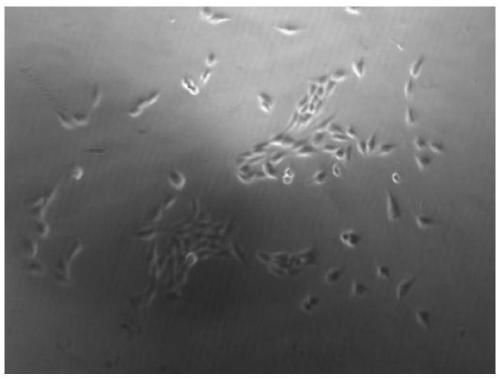
[0043] This embodiment discloses a method for isolating and culturing primary canine vascular endothelial cells, comprising the following steps:

[0044] (1) Get the puppy that just died in a car accident, clean up the blood and dirt on the corpse, shave the chest and abdomen in the aseptic operating room after wiping and disinfecting the chest and abdomen with tincture of iodine, and then wipe the tincture of iodine with 75% alcohol. Open the chest cavity, cut the aorta about 4cm away with sterile scissors, squeeze out the remaining congestion in the blood vessel with tweezers, soak it in 75% alcohol for 10 seconds, take it out and place it in a clean workbench;

[0045] (2) Soak the blood vessel with PBS solution containing 5% double antibody solution (penicillin-streptomycin mixed solution), carefully peel off the hoof tissue attached to the surface of the blood vessel, and then use sterile pointed forceps to insert one end of the blood vessel inward Lumen, slowly move the ...

Embodiment 2

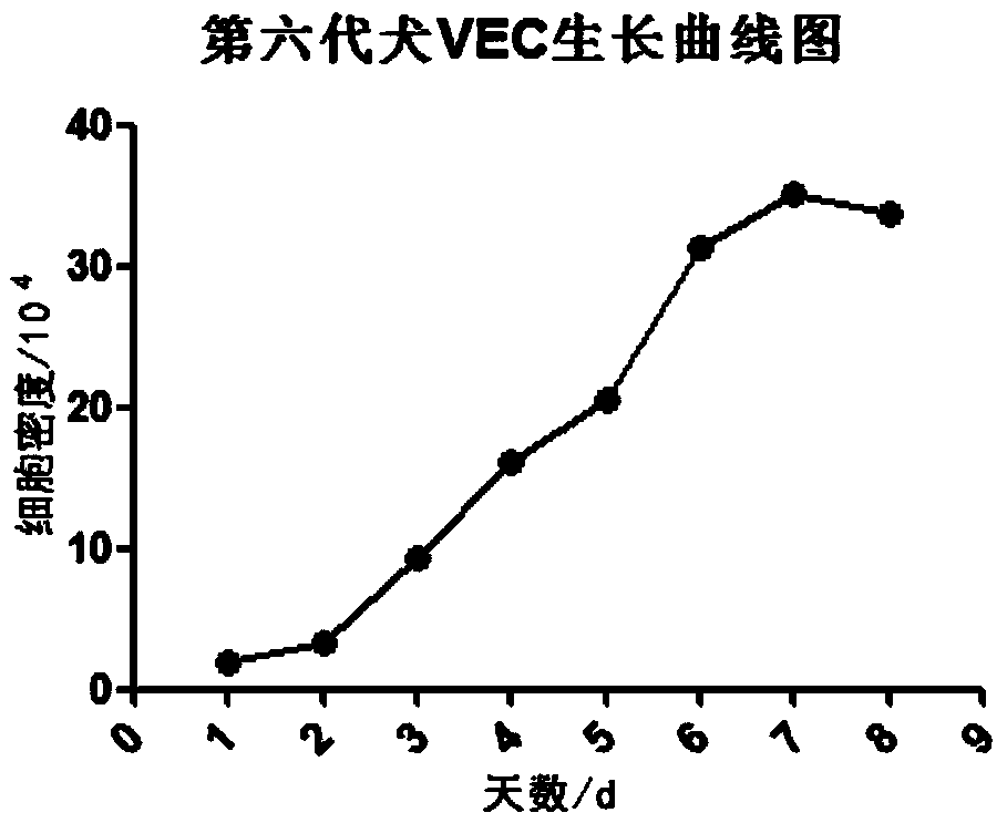
[0057] In this embodiment, the growth curve of canine vascular endothelial cells (VEC) is drawn, and the specific operations are as follows:

[0058](1) The primary canine vascular endothelial cells in Example 1 were subcultured to the sixth generation, and the well-grown vascular endothelial cells were taken and digested with 0.25% trypsin to make a cell suspension;

[0059] (2) Dilute the cells to 2×10 4 / mL inoculated into 24-well plate, 0.5mL per well, placed in 5% CO 2 , cultured in a 37°C incubator;

[0060] (3) Randomly extract cells from 3 wells at the same time every day, digest them with trypsin to make a suspension, count them under a microscope, and count three times in each well to get the average value;

[0061] (4) Continuous counting for eight days, according to the counting results, with the cell density as the ordinate (unit / mL) and the time as the abscissa, draw as follows figure 2 The growth curve graph shown. From figure 2 It can be seen that the pr...

Embodiment 3

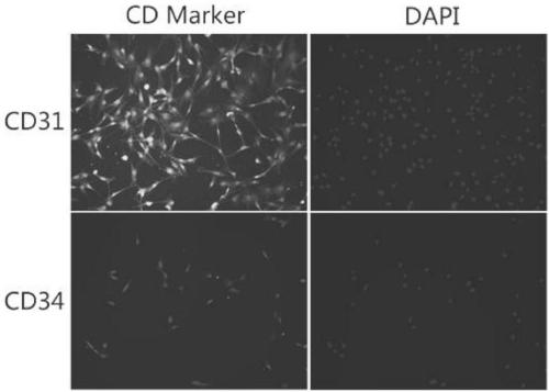
[0063] In this example, an immunofluorescence identification experiment was performed on the sixth-generation canine vascular endothelial cells, and the specific steps were as follows:

[0064] (1) The primary canine vascular endothelial cells in Example 1 are subcultured to the sixth generation, and the well-grown vascular endothelial cells are taken, seeded on a gelatin-coated culture plate, and the cells are adhered to the wall for 12 hours, discarded The culture medium was washed twice with PBS solution containing 5% FBS (fetal bovine serum);

[0065] (2) Add 4% paraformaldehyde containing 0.1% Triton×100 to fix at room temperature for 5 minutes, remove the fixative, and wash 3 times with PBS solution containing 5% FBS;

[0066] (4) Add blocking solution (PBS containing 10% FBS), block at 37°C for 1 hour, remove the blocking solution, and wash 3 times with PBS solution containing 5% FBS;

[0067] (5) Add the primary antibodies of CD31 (purchased from Bioss, Cat. No. bs-09...


PUM


Abstract

The invention relates to the field of biology, in particular to an isolated culture method for primary canine vascular endothelial cells. The isolated culture method comprises the following steps: S1,taking canine aorta; S2, turning over the canine aorta and cleaning; S3, obtaining vascular endothelinal surface single-layer cells; and S4, obtaining the primary canine vascular endothelial cells and performing in-vitro culture. The isolated culture method disclosed by the invention has the advantages that: operations are simple, repeatability is good and experimental time is short; the vascularendothelial cells are derived from canine aorta dead of a car accident, so that ethical arguments are avoided; cells are scraped through a mechanical method, and are combined with a collagenase digestion method, so that a great number of vascular endothelial cells with strong multiplication capacity can be obtained; the obtained vascular endothelial cells are high in purity, and are very low in cell pollution probability. A turnover operation success rate is nearly 100%; and connective tissues adhered on the vascular surfaces are stripped off, so that the pollution chance of other cells suchas fibroblast is greatly reduced.

Description

technical field [0001] The invention relates to the field of biology, in particular to a method for isolating and culturing primary canine vascular endothelial cells. Background technique [0002] In today's high incidence of cardiovascular and cerebrovascular diseases, the establishment of disease models and drug development are particularly important. Vascular endothelial cells, as the most suitable model cells for cardiovascular and cerebrovascular diseases, have the characteristics of difficult separation and low purity, which to some extent restricts the development of cardiovascular and cerebrovascular disease drug research and development. Although there are many vascular endothelial cell lines on the market, they have undergone certain mutations compared with primary cells, and are quite different from vascular endothelial cells in the physiological environment in vivo. As the most popular companion pet, dogs are also troubled by veterinarians and owners due to thei...

Claims


Application Information

Patent Timeline
Patent Type & Authority Applications(China)
IPC IPC(8): C12N5/071
CPCC12N5/069C12N2509/00
Inventor 詹小舒王丙云罗冬章罗惠娜陈胜锋陈志胜刘璨颖白银山
Owner FOSHAN UNIVERSITY
Why Eureka
Try Eureka