A method for preparing 3D cartilage organoid block

An organoid, three-dimensional technology, used in biochemical equipment and methods, medical preparations containing active ingredients, pharmaceutical formulations, etc., can solve problems such as difficult and difficult conventional technology, preparation of therapeutic agents, etc. Compatibility, the effect of increasing biocompatibility

Active Publication Date: 2019-01-04
XCELL THERAPEUTICS INC
View PDF10 Cites 2 Cited by
  • Summary
  • Abstract
  • Description
  • Claims
  • Application Information

AI Technical Summary

Problems solved by technology

Although cell population culture techniques using 96 wells have been developed, which contain one spheroid in each single well, it is difficult to structurally form a tissue where the spheroids fuse with each other
It is not possible to prepare cartilage tissue from biomaterials such as hydrogels, making it difficult to prepare therapeutic agents using conventional techniques

Method used


Image

  • A method for preparing 3D cartilage organoid block
  • A method for preparing 3D cartilage organoid block
  • A method for preparing 3D cartilage organoid block

Examples


Embodiment 1

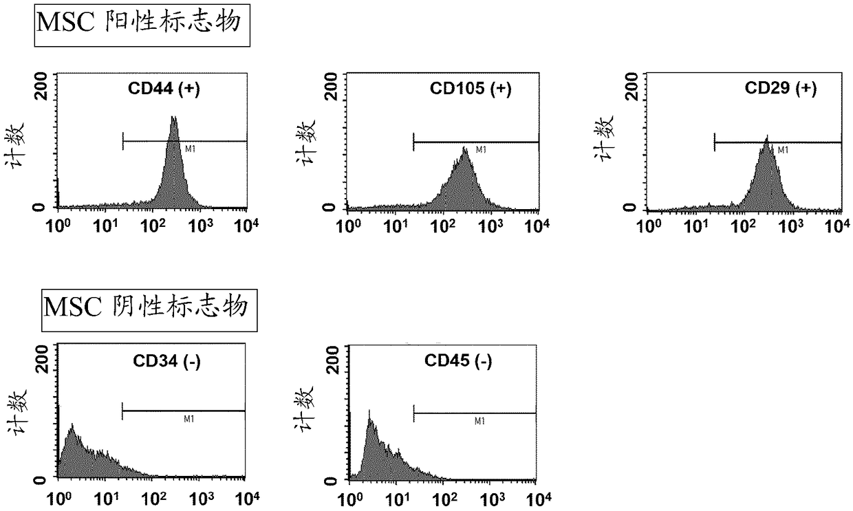
[0032] Example 1. Isolation, Incubation and Stemness Evaluation of Human Mesenchymal Stem Cells

[0033] Adipose tissue was randomly cut into small pieces, and the obtained pieces were washed three times with phosphate-buffered saline (PBS) (Sigma, St. Louis, MO). Then, place small pieces of adipose tissue into a 50 ml conical tube. Add PBS to the tube and stir, then centrifuge. The liquid was discarded, Dubeck's modified Eagle's medium (DMEM) was added to a volume of 50 ml, and the mixture was allowed to react at 37° C. for 90 minutes. After centrifugation at 2,000 rpm for 10 minutes, undissolved adipose tissue suspended in the upper layer was removed. Then, wash with DMEM, centrifuge and remove repeatedly. Separated adipose-derived stem cells were incubated with serum-free stem cell medium (chemically defined medium) at 37°C, 5% CO 2 Incubate and proliferate in an incubator. DMEM containing 10% FBS can be used for proliferation. Proliferating mesenchymal stem cells w...

Embodiment 2

[0034] Example 2, Evaluation of Mesenchymal Stem Cells Differentiation to Cartilage

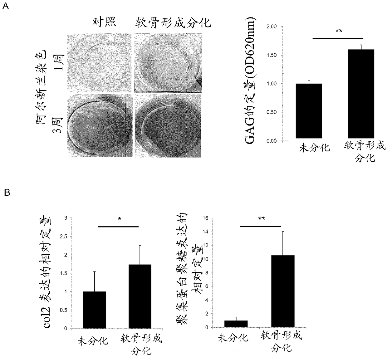
[0035] Stem cells obtained in 1x10 4 cells / cm 2 Inoculate and inoculate at 37 °C in 5% CO 2 The cells were cultured in an incubator, and the cells were treated with chondrogenic differentiation medium every 2 days in order to differentiate the mesenchymal stem cells into chondrocytes. Chondrogenic differentiation medium contained 50 μg / ml ascorbic acid 2-phosphate, 100 nM dexamethasone, 1% ITS and 10 ng / ml TGF-β1. The level of GAG matrix formation was fixed by treating cells in two-dimensional cartilage plates with 10% formaldehyde for 30 minutes, then cells were treated with 3% acetic acid solution for 3 minutes, followed by staining with 500 μl of Alcian blue solution (pH 2.5) for 30 minutes. Stained samples were washed several times with distilled water and examined microscopically. To obtain quantitative GAG ​​values, Alcian blue-stained plates were treated with 3% acetic acid for 1...

Embodiment 3

[0036] Example 3. Cartilage stimulation using real-time PCR

[0037] To analyze gene modification between undifferentiated stem cells and chondrogenic differentiated cells, the expression of chondrogenic differentiation genes was assessed. For this purpose, ColII and aggrecan were used as gene markers related to chondrocytes, and GAPDH was used as a housekeeping gene. Real-time PCR was performed as follows. That is, cells obtained from each group were washed with PBS, and then they were collected with trypsin-EDTA, and RNA was extracted by the TRIzol (Life Technologies, Inc. Grand Island, NY) method. 1 μg of extracted RNA was used to prepare cDNA and study changes in gene expression. Primer sets and corresponding differentiation markers are shown in Table 1 below.

[0038] Table 1

[0039]

[0040] *NCBI accession number

[0041] The results of the above real-time PCR are shown in figure 2 C. After chondrogenic differentiation, cartilage differentiation indicator ...


PUM


Abstract

The present invention is to provide a 3D cartilage organoid block prepared by differentiating mesenchymal stem cells into 3D spheroid cartilage tissues, a basic unit for the 3D cartilage spheroid block. The inventors found that both the amount of GAG matrix and the expression of the collagen type2 increased. Therefore, the method of this invention provides clinically applicable cartilage tissues by effectively enhancing the function of the cartilage differentiation constructs according to 2D culture. The 3D cartilage organoid block can be usefully applied to the area, such as, articular cartilage regeneration and plastic aurgery, where cartilage tissues restoration is required.

Description

technical field [0001] The present invention relates to the preparation of artificial tissues. The present invention directly relates to methods for preparing artificial cartilage tissue in vitro. Background technique [0002] Aging and increased social and physical activity lead to an increase in the number of patients suffering from cartilage damage, and thus the need to develop medical techniques to restore damaged cartilage is also increasing. However, since cartilage tissue has no blood vessels and the mobility of chondrocytes is limited by the extracellular matrix (ECM), it is difficult to easily regenerate damaged cartilage. [0003] Among the current methods of treating damaged cartilage, artificial joint replacements have the advantage of relieving pain and allowing patients to walk naturally when the procedure is successfully completed. Since the technology related to artificial joint replacement has improved greatly recently, artificial joint replacement surgery...

Claims


Application Information

Patent Timeline
Patent Type & Authority Applications(China)
IPC IPC(8): A61L27/38A61K35/32A61K35/28C12N5/077
CPCA61K35/32A61L27/3834A61L27/3852A61L27/3895A61K35/28C12N5/0671A61L2430/06C12N2506/13C12N5/0655C12N2506/1346C12N2513/00
Inventor 申永基朴相奎金永德安钟赞朴炳俊李锡逸
Owner XCELL THERAPEUTICS INC
Why Eureka
Try Eureka