
Visual positioning guide wire

A guide wire and positioning sensor technology, which is applied in the field of medical devices, can solve the problems of high cost and easy breakage of glass fiber filaments, and achieve the effects of low manufacturing cost, avoiding cross-infection, and clear imaging

Pending Publication Date: 2019-05-24
SHANGHAI LINCHAO MEDICAL INSTR
View PDF12 Cites 1 Cited by
  • Summary
  • Abstract
  • Description
  • Claims
  • Application Information

AI Technical Summary

Problems solved by technology

[0015] The technical problem to be solved by the present invention is to provide a visual positioning guide wire, which overcomes the defects of the prior art, solves the last blind area in the operation of peripheral lung lesions, and makes the guide wire visual and locatable. After reaching the target area through navigation , so that doctors can look directly at the lesion; it solves the problems of high cost, easy breakage of glass fiber filaments, reliability problems and imaging quality problems of existing optical fiber imaging catheters

Method used


Image

  • Visual positioning guide wire
  • Visual positioning guide wire
  • Visual positioning guide wire

Examples


Embodiment Construction

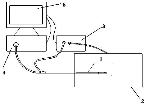
[0041] Such as figure 1 As shown, the visual positioning guide wire 1 of the present invention is connected to the image processing system 4 and the navigation system 3 respectively, and the output of one electromagnetic positioning signal of the visual positioning guide wire 1 (that is, the signal line of the electromagnetic positioning sensor) can be directly connected to the navigation system 3 superior. The image processing system 4 is connected with the navigation system 3, and its main purpose is to simultaneously display the endoscopic image and navigation positioning information in one screen, and switch in real time. This feature is available in current navigation software. Therefore, the current practice is that the image processing system 4 has a video output function, and the video signal is output to the navigation system 3, and then displayed by the navigation system 3 together; in the later stage, the endoscopic image and the navigation image can be superimpose...


PUM


Abstract

The invention discloses a visual positioning guide wire. An end of the visual positioning guide wire is provided with a positioning sensor; the front end of the positioning sensor is provided with a visual device; the visual device includes a CMOS (complementary metal oxide semiconductor) sensor, an imaging lens and an optical fiber. The defects of the prior art are overcome; the last blind area is eliminated for peripheral lung disease surgery is eliminated, and the guide wire is visually positionable; after the guide wire reaches a target region through navigation, a doctor can see the focus; the problems are solved that an existing fiber imaging catheter has high cost, the glass fiber yarns break easily, the reliability is poor and the imaging quality is poor.

Description

technical field [0001] The present invention relates to a medical device, in particular to a medical guide wire, or a disposable endoscope, in particular to a visual positioning guide wire. Background technique [0002] The diagnosis of peripheral pulmonary lesions has always been a difficult problem for clinicians. Commonly used examination methods include routine bronchoscopy, transthoracic lung biopsy, endobronchial radial ultrasound, and diagnostic surgery. However, most peripheral pulmonary lesions cannot be directly observed or only some indirect signs can be seen under bronchoscopy, and the diagnostic rate of peripheral pulmonary lesions by bronchoscopy is affected by the size of the lesion. The smaller the lesion, the lower the diagnostic rate. [0003] After 2000, a new technique for the diagnosis of peripheral lung lesions - electromagnetic navigation bronchoscopy (ENB for short) came out. ENB technology integrates the advantages of simulated bronchoscope and fle...

Claims


Application Information

Patent Timeline
Patent Type & Authority Applications(China)
IPC IPC(8): A61B1/267A61B1/07A61B1/05
Inventor 高科全懿
Owner SHANGHAI LINCHAO MEDICAL INSTR
Learn More