Near-infrared II-region fluorescent quantum dot cytomembrane marking system and marking method and application

A fluorescent quantum dot and labeling system technology, applied in the field of nano-biological materials, can solve the problems of low tissue penetration depth, easy quenching of fluorescent probes, high spontaneous background fluorescence, etc., to achieve good tissue penetration depth, good labeling and The effect of related function research and simplified experimental operation

Inactive Publication Date: 2019-06-07
SUZHOU INST OF NANO TECH & NANO BIONICS CHINESE ACEDEMY OF SCI
View PDF3 Cites 1 Cited by
  • Summary
  • Abstract
  • Description
  • Claims
  • Application Information

AI Technical Summary

Problems solved by technology

Currently commercially available cell membrane labeling probes are mainly concentrated in the 400-900nm band, such as the DiI cell membrane orange-red fluorescent probe, which can emit orange-red fluorescence after being excited, but this kind of fluorescent probe has a high quenching Extinction constant, resulting in low photon intensity and short excited state lifetime, which makes fluorescent probes easy to quench, which is not conducive to long-term observation in the process of cell membrane research; in addition, some fluorescent probes have excitation wavelengths in the ultraviolet region , its high excitation energy leads to greater damage to the cells after long-term irradiation, which is not conducive to the research
At the same time, traditional fluorescent probes (400-900nm) have the disadvantages of strong absorption, high scattering and high spontaneous background fluorescence in living tissues, which face low tissue penetration depth (<5 mm) and low spatial resolution in in vivo imaging research. Challenges such as low (about 1000 microns) and low time resolution (about 1000 milliseconds) seriously restrict its wide application

Method used


Image

  • Near-infrared II-region fluorescent quantum dot cytomembrane marking system and marking method and application
  • Near-infrared II-region fluorescent quantum dot cytomembrane marking system and marking method and application
  • Near-infrared II-region fluorescent quantum dot cytomembrane marking system and marking method and application

Examples


preparation example Construction

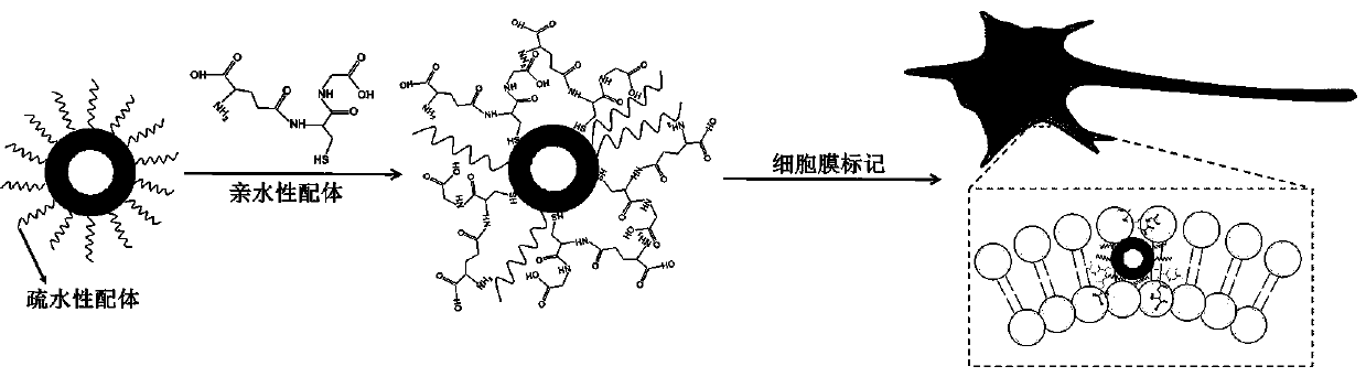
[0035] Another aspect of the embodiments of the present invention also provides a preparation method for the aforementioned near-infrared II region fluorescent quantum dot cell membrane labeling system, which includes: making hydrophobic ligands and hydrophilic ligands respectively modified and connected to the near-infrared II region fluorescent quantum dots Dot the surface to obtain the near-infrared II region fluorescent quantum dot cell membrane labeling system.

[0036]In some embodiments, the preparation method specifically includes:

[0037] The hydrophobic ligand is modified and connected to the surface of the fluorescent quantum dot in the near-infrared II region to form a hydrophobic ligand quantum dot;

[0038] The hydrophilic ligand, the hydrophobic ligand quantum dot and the solvent are uniformly mixed, stirred, and centrifuged to obtain the near-infrared II region fluorescent quantum dot cell membrane labeling system.

[0039] In some more specific implementatio...

Embodiment 1

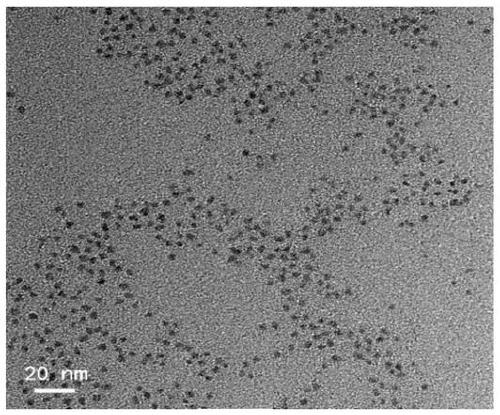
[0056] Hydrophilic GSH-DT-Ag 2 The specific preparation method of S quantum dots:

[0057] Take a clean and dry glass bottle, add chloroform and deionized water to it, and add DT-Ag according to a certain molar concentration ratio 2 S and glutathione (GSH), after mixing by magnetic stirring, continue to stir under the condition of avoiding light, and finally take the upper water phase and the liquid at the oil-water interface, discard the upper water phase by centrifugation, and wash once with deionized water Obtained GSH-DT-Ag after centrifugation and resuspension 2 S water-soluble quantum dots. GSH-DT-Ag prepared 2 The TEM characterization data of S hydrophilic quantum dots are as follows: figure 2 As shown, the average particle size is about 2 nm.

Embodiment 2

[0059] Hydrophilic DHLA-DT-Ag 2 The specific preparation method of S quantum dots:

[0060] Take a clean and dry glass bottle, add chloroform and deionized water to it, and add DT-Ag according to a certain molar concentration ratio 2 S and dihydrolipoic acid (DHLA), after mixing by magnetic stirring, continue to stir under the condition of avoiding light, and finally take the upper water phase and the liquid at the oil-water interface, discard the upper water phase by centrifugation, and wash once with deionized water DHLA-DT-Ag obtained after centrifugation and resuspension 2 S water-soluble quantum dots. The preparation method of other hydrophilic hydrophilic / phobic double ligand quantum dots is similar.


PUM

PropertyMeasurementUnit
Centrifugal forceaaaaaaaaaa

Abstract

The invention discloses a near-infrared II-region fluorescent quantum dot cytomembrane marking system and marking method and application. The near-infrared II-region fluorescent quantum dot cytomembrane marking system comprises near-infrared II-region fluorescent quantum dots and hydrophilic ligands and hydrophobic ligands which are connected with the surfaces of the near-infrared II-region fluorescent quantum dots in a modifying mode. The marking method comprises the steps of making to-be-marked cells and the near-infrared II-region fluorescent quantum dot cytomembrane marking system mixed toform a marking system, and then conducting co-incubation on the marking system to make the near-infrared II-region fluorescent quantum dot cytomembrane marking system embedded into lipid bilayers ofcytomembrane to achieve cytomembrane marking. The near-infrared II-region fluorescent quantum dot cytomembrane marking system can be successfully embedded into the lipid bilayers of the cytomembrane to achieve cytomembrane marking and fluorescence imaging, so that the near-infrared II-region fluorescent quantum dot cytomembrane marking system has large application potential and values for markinglive cells.

Description

technical field [0001] The present invention relates to a cell membrane labeling system, in particular to a near-infrared II region fluorescent quantum dot cell membrane labeling system, a labeling method, and the application of high-affinity labeling and cell membrane imaging for cell membranes, and can be used as a favorable tool for cell membrane labeling in conjunction with cell membranes The use in function-related research belongs to the technical field of nanobiological materials. Background technique [0002] The cell membrane is composed of phospholipid bilayer molecules as the basic unit repeating, that is, the phospholipid bilayer, on which various types of membrane proteins and sugars and glycolipids bound to membrane proteins are embedded, which are involved in various cellular processes, such as Material transport, cell signaling, cell adhesion, ion conduction, and more. In order to better understand the function and function of the cell membrane and its influ...

Claims


Application Information

Patent Timeline
IPC IPC(8): C09K11/58C09K11/02B82Y20/00B82Y30/00B82Y40/00G01N21/64
Inventor 王强斌彭钊陈光村张叶俊
Owner SUZHOU INST OF NANO TECH & NANO BIONICS CHINESE ACEDEMY OF SCI
Why Eureka
Try Eureka