
Human cervix uterus epithelial cell separation and culture method

An epithelial cell and culture method technology, applied in the field of separation and culture of human cervical epithelial cells, can solve the problems of immaturity of cervical epithelial cells in vitro, maintain cell physiological activity and proliferation ability, promote cell growth and proliferation, promote The effect of growth and division

Pending Publication Date: 2019-07-05
北京昱龙摩尔国际生物医学研究院
View PDF2 Cites 0 Cited by
  • Summary
  • Abstract
  • Description
  • Claims
  • Application Information

AI Technical Summary

Problems solved by technology

However, the in vitro culture of cervical epithelial cells is still immature

Method used


Image

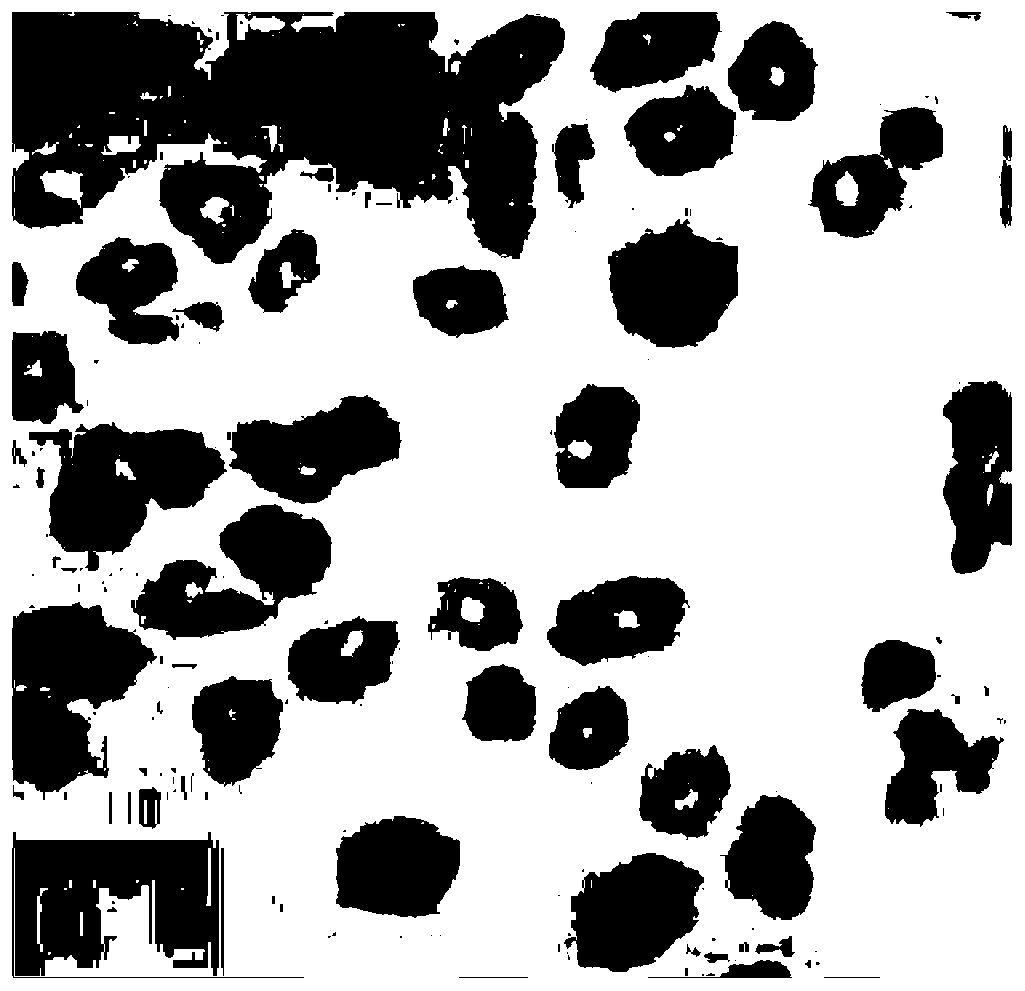
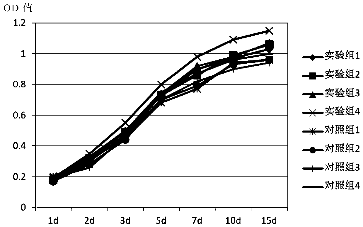
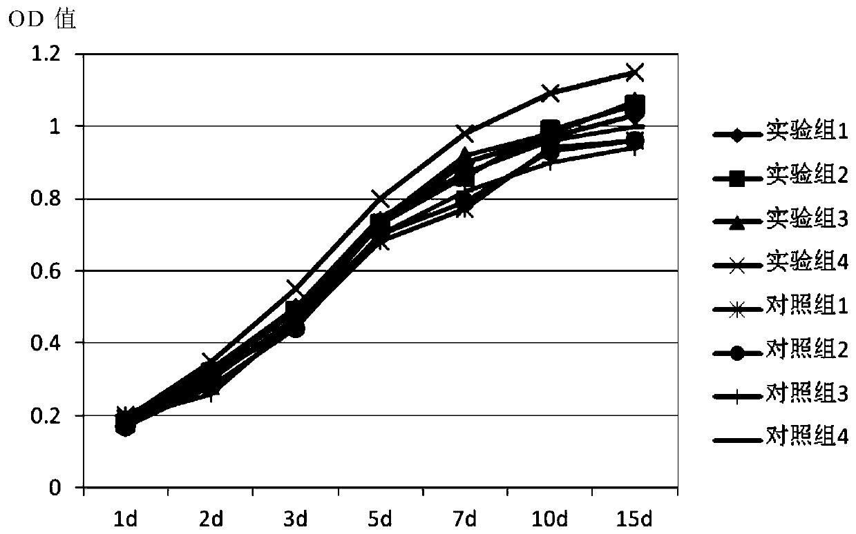
  • Human cervix uterus epithelial cell separation and culture method
  • Human cervix uterus epithelial cell separation and culture method
  • Human cervix uterus epithelial cell separation and culture method

Examples


Embodiment 1

[0050] A method for isolating and culturing human cervical epithelial cells, comprising the steps of:

[0051] S1: Sample collection and separation: collect biopsy specimens, rinse with PBS, remove fibrous tissue, obtain cervical epithelial tissue, and cut into 2mm tissue pieces for later use;

[0052] S2: washing and sedimenting the tissue block with PBS buffer solution, discarding the PBS and then repeating with the culture solution, discarding the culture solution, and keeping the tissue block for later use;

[0053] S3: Primary culture: Place the tissue pieces in a culture bottle for the first stage of adherent culture, then add the primary culture medium for the second stage of adherent culture to obtain primary cells;

[0054] S4: Subculture: after the primary cells were digested, 10 4 The concentration of the cells was inoculated, and the irradiated 3T3 cells were used as the feeder layer, and the subculture medium was added for suspension culture to obtain the subcult...

Embodiment 2

[0056] A method for isolating and culturing human cervical epithelial cells, comprising the steps provided in Example 1, wherein the specific method of step S1 is as follows:

[0057] S1.1: Collect biopsy specimens and place them in preservation solution for later use;

[0058] S1.2: Take five 9cm petri dishes, add 15ml of PBS buffer to No. 1, No. 2 and No. 3 petri dishes respectively, add 5ml of PBS buffer to No. 4 petri dish, and do not add to No. 5 petri dish; After rinsing with PBS, put it into No. 1 petri dish for cleaning, take it out and put it into No. 2 petri dish, and use a scalpel to separate the fibrous tissue on the specimen, and retain the cervical epithelial tissue;

[0059] S1.3: Move the cervical epithelial tissue into No. 3 Petri dish for cleaning, take it out, transfer it to No. 4 Petri dish, and chop it into small pieces; move the small piece into No. 5 Petri dish, continue to cut into 2mm tissue pieces, and wash with PBS. Clean up.

[0060] The specific ...

Embodiment 3

[0074] A method for isolating and culturing human cervical epithelial cells, comprising the steps provided in Example 2, wherein the primary culture medium used is the Leibovitz L-15 medium added with the following components:

[0075] 10% fetal bovine serum, 0.5 μg / ml hydrocortisone, 50 U / ml penicillin, 50 U / ml streptomycin and 5 ng / ml EGF.


PUM


Abstract

The invention provides a human cervix uterus epithelial cell separation and culture method. The method has the advantages that a collected biopsy sample is preserved in a special preservation liquid,so that cell physiological activity and multiplication capacity can be kept effectively; cell growth factors are added into a primary culture medium, so that cell activity can be increased effectively, cell growth can be promoted, and high-quality primary cells; allantoin, creatine and vitexin are added into a passage culture medium, so that cell activity and division ability can be increased effectively, and cell growth and proliferation are promoted; 3T3 cells are used as the nourishing layer, a good growth substrate can be provided for the primary cervix uterus epithelial cells, the growthand division of the cervix uterus epithelial cells are promoted effectively, and the culture effect is increased greatly; by the method, the activity and proliferation speed of the cervix uterus epithelial cell can be evidently increased, and high-quality experiment materials can be provided for related research projects.

Description

technical field [0001] The invention belongs to the technical field of tissue engineering, in particular to a method for separating and culturing human cervical epithelial cells. Background technique [0002] Cervical cancer is the malignant tumor with the highest incidence rate of female reproductive system in developing countries, and its main cause is the persistent infection of high-risk human papillomavirus (human papilloma virus, HPV) in cervical epithelial cells. The cervical cancer cell lines commonly used in current research (such as HeLa cell lines, etc.) are all in an integrated state, and it is difficult to reflect the cell status in the early stage of HPV infection. Freshly isolated primary cells retain their original biological genetic characteristics and can reflect the general characteristics of cell growth in vivo, so they are suitable for research on drugs and cancer treatment. The establishment of in vitro cell models of normal cervical epithelial cells a...

Claims


Application Information

Patent Timeline
Patent Type & Authority Applications(China)
IPC IPC(8): C12N5/071
CPCC12N5/0682C12N5/0625C12N2502/02C12N2501/11C12N2501/39C12N2500/12C12N2500/14C12N2501/999C12N2509/00
Inventor 张晓南吴芳春吴刘兵陈虎张斌侍晓云
Owner 北京昱龙摩尔国际生物医学研究院
Learn More