Growth factor sustained release microsphere, tissue engineering cartilage composite stent and preparation method

A growth factor and slow-release microsphere technology, applied in the field of medical materials, can solve the problems of difficulty in realizing self-repair and regeneration, aggravation, and low self-repair and regeneration ability of cartilage tissue, and achieve the effect of improving the effect of regeneration and repair.

Active Publication Date: 2019-08-27
GENERAL HOSPITAL OF PLA
View PDF2 Cites 7 Cited by
  • Summary
  • Abstract
  • Description
  • Claims
  • Application Information

AI Technical Summary

Problems solved by technology

It is precisely because of the histological characteristics of cartilage, the composition of biochemical components, and the characteristics of low metabolism and low proliferation of chondrocytes that limit the proliferation response and migration of chondrocyte...

Method used


Image

  • Growth factor sustained release microsphere, tissue engineering cartilage composite stent and preparation method
  • Growth factor sustained release microsphere, tissue engineering cartilage composite stent and preparation method
  • Growth factor sustained release microsphere, tissue engineering cartilage composite stent and preparation method

Examples


preparation example Construction

[0056] The second aspect of the embodiments of the present invention provides a preparation method of growth factor sustained-release microspheres, through which the above-mentioned growth factor sustained-release microspheres can be prepared. The preparation method comprises the following steps:

[0057] S10, providing an aqueous rhTGF-β3 solution with a concentration of 1 μg / mL˜100 μg / mL.

[0058] In step S10, rhTGF-β3 may be dissolved in deionized water to prepare an aqueous solution of rhTGF-β3 with a concentration of 1 μg / mL-100 μg / mL as the internal water phase w 1 .

[0059] S20, providing a PLGA oil phase solution with a concentration of 20 mg / mL-25 mg / mL.

[0060] In step S20, PLGA may be dissolved in dichloromethane (DCM) to prepare a PLGA oil phase solution with a concentration of 20 mg / mL-25 mg / mL as the oil phase o. The oil phase o can have a higher encapsulation efficiency for rhTGF-β3.

[0061] In step S20, the molecular weight of PLGA is preferably 1×10 5 ...

Embodiment 1

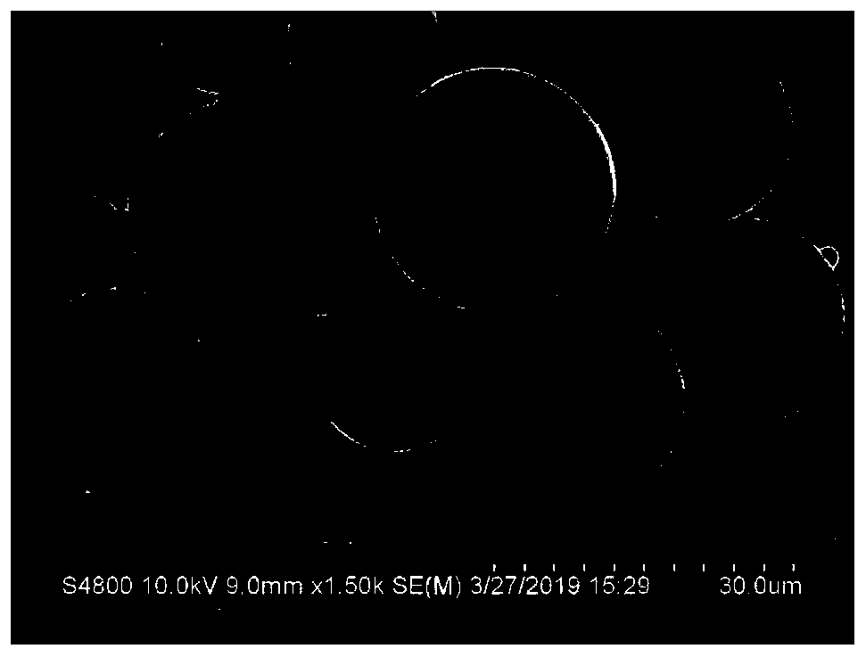
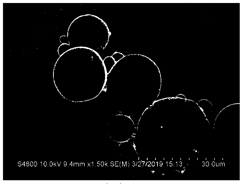
[0112] use w 1 / o / w 2 The growth factor sustained-release microspheres loaded with rhTGF-β3 in PLGA were prepared by emulsification-solvent evaporation method. The specific process was as follows: 2 μg of rhTGF-β3 was dissolved in 200 μL of deionized water to prepare an aqueous solution of rhTGF-β3 with a concentration of 10 μg / mL. Water phase w 1 . Dissolve polylactic-co-glycolic acid (PLGA) with a molecular weight of 102,000 Da in dichloromethane (DCM) to prepare a PLGA solution with a concentration of 25 mg / mL as the oil phase o. Polyvinyl alcohol (PVA) with a molecular weight of 67,000 Da was dissolved in deionized water to prepare a 1% PVA aqueous solution as the external water phase w 2 ; Dispense 200 μL of the inner aqueous phase w 1 Join in 2mL oily phase o, form primary emulsion (w 1 / o); then the primary emulsion (w 1 / o) Add 20mL external aqueous phase w 2 At room temperature (20°C-25°C), stir magnetically at 500rpm for 12 hours, volatilize and remove DCM to ...

Embodiment 2

[0114] The difference from Example 1 is that 5 μg of rhTGF-β3 was dissolved in 200 μL of deionized water to prepare a rhTGF-β3 aqueous solution with a concentration of 25 μg / mL as the internal water phase w 1 .


PUM

PropertyMeasurementUnit
Particle sizeaaaaaaaaaa
Heightaaaaaaaaaa
Widthaaaaaaaaaa

Abstract

The invention discloses a growth factor sustained release microsphere, a tissue engineering cartilage composite stent and a preparation method. The growth factor sustained release microsphere includesa polylactic acid-glycolic acid copolymer matrix material and recombinant human transforming growth factor beta 3 encapsulated in the matrix material, wherein the encapsulation amount of the recombinant human transforming growth factor beta 3 in the microsphere is 5 ng/mg to 200 ng/mg; the sustained release period of the microsphere is 28 days or above. The growth factor sustained release microsphere and the tissue engineering cartilage composite stent disclosed by the invention have higher regeneration and repair effects of defected cartilage tissues.

Description

technical field [0001] The invention belongs to the technical field of medical materials, and in particular relates to a growth factor slow-release microsphere, a tissue engineering cartilage composite scaffold and a preparation method. Background technique [0002] Cartilage is an important part of the skeletal system in humans or animals. Cartilage tissue is a highly specialized connective tissue, which is characterized by the lack of blood vessels, nerves and lymphatic vessels in the tissue. The extracellular matrix is ​​dense and solid. Chondrocytes are surrounded by a large number of cells. External matrix (such as collagen, fibers, proteins, polysaccharides, etc.) surrounded. It is precisely because of the histological characteristics of cartilage, the composition of biochemical components, and the characteristics of low metabolism and low proliferation of chondrocytes that limit the proliferation response and migration of chondrocytes to the damaged area, resulting in...

Claims


Application Information

Patent Timeline
IPC IPC(8): A61K9/52A61K38/18A61K47/34A61L27/18A61L27/22A61L27/52A61L27/54A61L27/58A61P19/08
CPCA61K38/1841A61K9/5031A61P19/08A61L27/227A61L27/18A61L27/54A61L27/52A61L27/58A61L2300/414A61L2300/622A61L2300/604A61L2430/06C08L89/00C08L67/04
Inventor 郭全义杨振卢世璧刘舒云眭翔黄靖香郭维民田广招孙志强查康康周建
Owner GENERAL HOSPITAL OF PLA
Why Eureka
Try Eureka