Tissue engineering bionic liver lobe structure based on 3D printing of living cells and preparation method thereof

A 3D printing and tissue engineering technology, applied in the field of biomedical engineering, can solve problems such as difficulty in achieving long-term survival of large-volume artificial tissues, difficulty in forming cell function integration, and support barriers

Active Publication Date: 2019-11-05
THE FIRST AFFILIATED HOSPITAL OF ARMY MEDICAL UNIV
View PDF6 Cites 7 Cited by
  • Summary
  • Abstract
  • Description
  • Claims
  • Application Information

AI Technical Summary

Problems solved by technology

For example, cells are seeded on a porous scaffold and soaked in the medium for culture, so that nutrients can reach the cells through the pores, but this method has certain limitations: as the cells grow, the pores in the scaffold will be filled by cells. The flow of nutrients is blocked, and the large-volume artificial tissue formed by this method is difficult to form the functional integration of cells in normal tissues due to the barrier of the scaffold material; in addition, some researchers use hydrogel materials that can induce blood vessel ingrowth However, this method still has the problem of deep support obstacles. When the blood vessels have not grown in...

Method used


Image

  • Tissue engineering bionic liver lobe structure based on 3D printing of living cells and preparation method thereof
  • Tissue engineering bionic liver lobe structure based on 3D printing of living cells and preparation method thereof
  • Tissue engineering bionic liver lobe structure based on 3D printing of living cells and preparation method thereof

Examples


Embodiment 1

[0038] Example 1 (Temperature-controlled dual-stage etching)

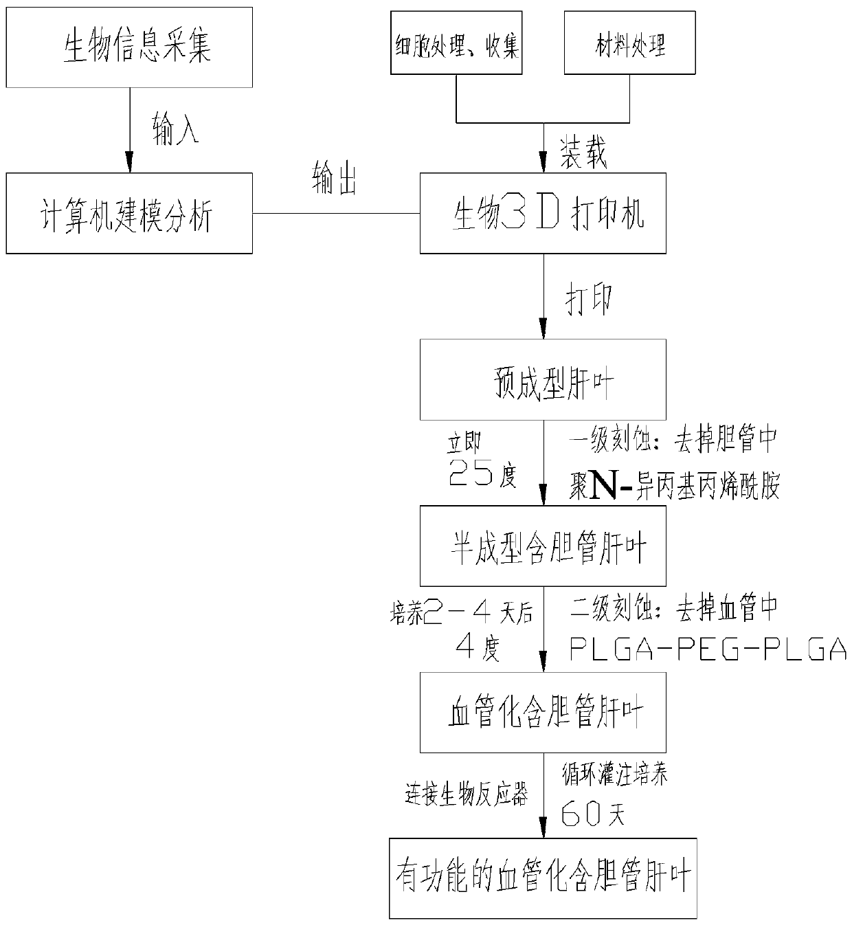
[0039] see figure 1 , this embodiment discloses a method for preparing a bionic liver lobe for tissue engineering based on living cell 3D printing, comprising the following steps:

[0040] 1. Biological information collection and modeling:

[0041] 1) Personalized collection of three-dimensional data of the internal and external structures of the liver lobe containing bile ducts and the blood circulation network of normal people through CT, nuclear magnetic resonance and micro three-dimensional scanning technology;

[0042] 2) Input the collected biological information into the computer software, and imitate the actual tissue appearance and microenvironment to express it as a multi-material, multi-scale geometric model. The two ends of the vascular network of the liver lobe can be designed as two main blood vessels entering and exiting for communication with The culture fluid delivery tubes are connected to form ...

Embodiment 2

[0062] Example 2 (temperature control, enzyme dual-stage etching)

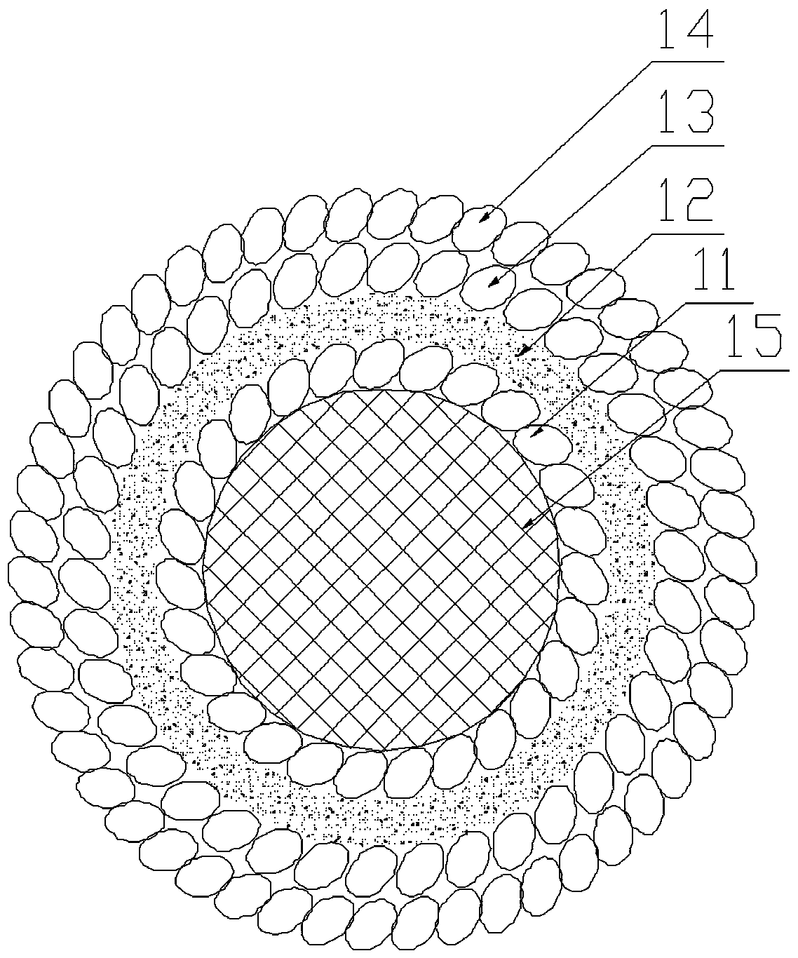
[0063] The first casting agent in this embodiment is fibrinogen and thrombin. Prepare fibrinogen, thrombin and fibrinase solutions respectively. The fibrinogen solution was prepared at a concentration of 100 mg / ml, and the thrombin and fibrinase solutions at a concentration of 1 U / ml, which were stored in a 4°C refrigerator before printing. The nozzle that sprays out cell-free fibrinogen and thrombin is maintained at 4°C without a UV light source. The nozzle for ejecting fibrinogen and thrombin is divided into two layers to separate the fibrinogen and thrombin, and the fibrinogen and thrombin are mixed together after ejection.

[0064] The second molding agent in this embodiment is poly-N-isopropylacrylamide. The prepared poly-N-isopropylacrylamide solution had a concentration of 100 mg / ml and stored it in a 4 °C refrigerator before printing to keep it in a liquid state. The nozzle spraying cell-free poly-...

Embodiment 3

[0068] Example 3 (Temperature control, chelation reaction dual-stage etching)

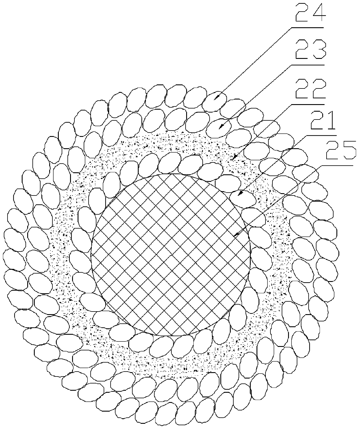
[0069] The first casting agent of this embodiment is sodium alginate and CaCl without cells 2 . Preparation of sodium alginate, CaCl 2 , Ethylenediaminetetraacetic acid (EDTA) solution. The prepared sodium alginate mass fraction is 10%, CaCl 2 The mass fraction is 0.5%. EDTA was dissolved in NaOH at a concentration of 100 mg / ml. Spray cell-free sodium alginate and CaCl 2 The spray head is kept at 4°C without UV light source. Which sprays sodium alginate and CaCl 2 The nozzle is divided into two layers, sodium alginate and CaCl 2 Separated, sodium alginate and CaCl after spraying 2 just mixed together.

[0070] The second molding agent in this embodiment is poly-N-isopropylacrylamide. The prepared poly-N-isopropylacrylamide solution had a concentration of 100 mg / ml and stored it in a 4 °C refrigerator before printing to keep it in a liquid state. The nozzle spraying cell-free poly-N-isop...


PUM

PropertyMeasurementUnit
Concentrationaaaaaaaaaa

Abstract

The invention discloses a tissue engineering bionic liver lobe structure based on 3D printing of living cells and a preparation method thereof. Firstly, biological information of human body liver lobes is collected, and inputted into a computer for high biomimetic modeling; then, material separated printing and material-cell mixed printing output are performed with a hydrogel material and cells for constructing liver, bile duct epithelium, blood vessel endothelium, smooth muscle and fibroblast and the like required for the human liver lobes by a biological 3D printer, wherein a hollow part inblood vessels and bile ducts is printed by a casting agent; after all the hydrogel materials are solidified, the casting agent in the blood vessels and bile ducts are removed by means of enzymes or chelating reactions or temperature control or illumination and the like, and a vascular system and a bile duct system in the artificial bile duct-containing liver lobes are constructed. The circulationfeeding of the vascular system of the bionic liver lobes and gas exchange are achieved by connection of main blood vessels at two ends of the vascular system and a delivery pipe of a culture solution.The long-term survival and biological function of the bionic liver lobes in vitro and in vivo are achieved.

Description

technical field [0001] The invention belongs to biomedical engineering, and particularly relates to a tissue engineering bionic liver lobe structure based on living cell 3D printing and a preparation method, that is, to prepare an artificial vascularized bile duct that can survive for a long time and has biological functions by adopting the method of biological 3D printing liver lobe. Background technique [0002] Existing artificial tissues are usually prepared by in vitro three-dimensional culture of cells, and only small-volume artificial tissues can be prepared. When the thickness of the artificial tissue exceeds 2 mm, it will cause deep tissue supply barriers, making it difficult for the artificial tissue to survive for a long time. In order to solve the problem of deep support barriers, researchers at home and abroad have carried out various explorations. For example, cells are seeded on porous scaffolds and soaked in medium for culture, so that nutrients can reach t...

Claims


Application Information

Patent Timeline
IPC IPC(8): A61F2/02A61L27/16A61L27/18A61L27/20A61L27/22A61L27/24A61L27/38A61L27/50A61L27/52A61L27/58B29C64/112B33Y10/00B33Y70/00B33Y80/00
CPCA61F2/022A61L27/16A61L27/18A61L27/20A61L27/222A61L27/227A61L27/24A61L27/38A61L27/3839A61L27/50A61L27/52A61L27/58B33Y10/00B33Y70/00B33Y80/00B29C64/112
Inventor 周强叶吉星甘翼搏李培王利元欧阳斌涂兵
Owner THE FIRST AFFILIATED HOSPITAL OF ARMY MEDICAL UNIV
Why Eureka
Try Eureka