A preparation method of self-beautifying biological material and self-beautifying biological material
A biomaterial and autologous technology, applied in the direction of prosthesis, tissue regeneration, animal cells, etc., can solve the problems of low purity and biological activity, long preparation cycle, and poor curative effect of biomaterials, so as to solve immune rejection and make up for the inadequacy of repair. Excellent, complete tissue compatibility
- Summary
- Abstract
- Description
- Claims
- Application Information
AI Technical Summary
Problems solved by technology
Method used
Image
Examples
Embodiment 1
[0041] This embodiment provides a method for making an autologous cosmetic biological material, comprising the following steps:
[0042] 1. Tissue collection: Under sterile conditions, use a skin sampler (skin trephine) to collect circular skin tissue with a diameter of 2-4mm behind the ear. Among them, the skin tissue behind the ear is a complete skin tissue, including epidermis, dermis, and subcutaneous tissue. Wash samples with phosphate buffered saline containing antibiotics and store in liquid nitrogen;
[0043] 2. Fibroblast isolation culture and primary (P0) cell culture:
[0044]Take out the tissue, wash it 1-2 times with DMEM (Dulbecco's Modified Eagle Medium) medium containing antibiotics after thawing, then use 75% alcohol to sterilize for a short time, and then wash it with phosphate buffer saline without calcium and magnesium ions. In this embodiment, epithelial cells and keratinocytes are removed by using a special culture medium for fibroblasts and the digesti...
Embodiment 2
[0074] This embodiment provides a method for making an autologous beauty biomaterial, the manufacturing steps of which are basically the same as those in Embodiment 1, the difference being that:
[0075] In fibroblast isolation culture and primary (P0) cell culture:
[0076] Take out the tissue, wash it 1-2 times with DMEM (Dulbecco's Modified Eagle Medium) medium containing antibiotics after thawing, then use 75% alcohol to sterilize for a short time, and then wash it with phosphate buffer saline without calcium and magnesium ions. Purify fibroblasts by removing epithelial cells and keratinocytes with fibroblast-specific culture medium and by limiting digestion time. Place the tissue in a composition containing collagenase and dispase for overnight digestion. After overnight digestion, remove the epidermis of the tissue block, then cut the tissue into pieces and move it to a special medium for culture for 20 days. When the degree of cell confluence reaches At 80%, the cells ...
Embodiment 3
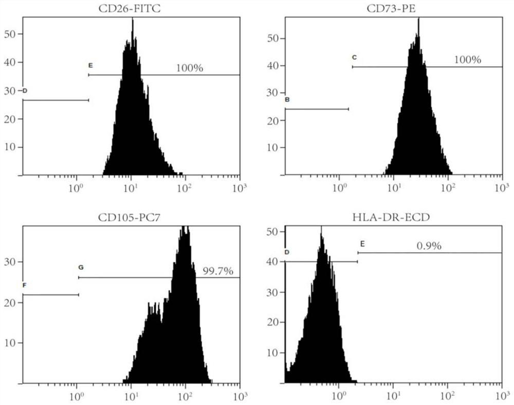
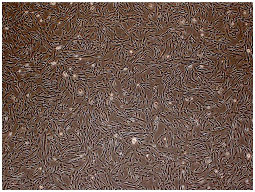
[0095] This example provides a self-cosmetic biological material prepared by the method for making the self-cosmetic biomaterial described in Example 1. The self-cosmetic biomaterial is composed of fibroblasts, physiological saline, and plasma rich in autologous platelets. , cell growth factors, collagen.
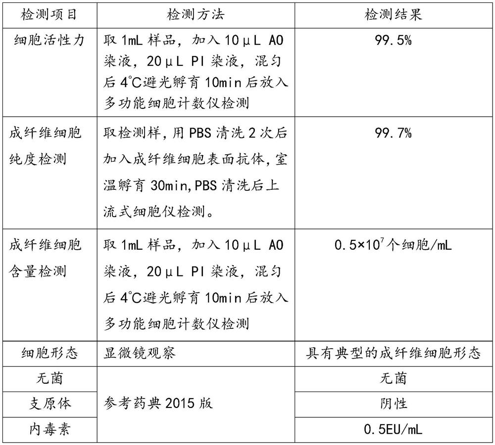
[0096] The number of third-generation fibroblasts is 0.5*10 7 indivual.
[0097] The purity of the third passage fibroblasts was 99.7%.
[0098] The viability of the third passage fibroblasts was 99.5%.
[0099] The content of autologous plasma in 1ml of autologous beauty biomaterial is 0.8mL.
[0100] The content of physiological saline in 1ml of the self-cosmetic biological material is about 0.2mL.
[0101] The cell growth factors in the self-cosmetic biological material include fibroblast growth factor, epidermal growth factor, platelet-derived growth factor, transforming growth factor, vascular endothelial cell growth factor and insulin-like growth factor.
[0102]...
PUM
 Abstract
Description
Claims
Application Information
 - R&D
- Intellectual Property
- Life Sciences
- Materials
- Tech Scout
- Unparalleled Data Quality
- Higher Quality Content
- 60% Fewer Hallucinations
Browse by: Latest US Patents, China's latest patents, Technical Efficacy Thesaurus, Application Domain, Technology Topic, Popular Technical Reports.
© 2025 PatSnap. All rights reserved.Legal|Privacy policy|Modern Slavery Act Transparency Statement|Sitemap|About US| Contact US: help@patsnap.com


