Method for constructing cartilage patch stent based on polycaprolactone electrostatic spinning 3D printing and device thereof

An electrospinning and polycaprolactone technology, applied in the medical field, can solve problems such as difficult stent area, thickness modification, and inability to adjust the internal structure design of the stent, so as to improve biomechanical properties, superior biomechanical properties, and broaden applications range effect

Inactive Publication Date: 2020-03-06
SHANGHAI PULMONARY HOSPITAL
View PDF4 Cites 2 Cited by
  • Summary
  • Abstract
  • Description
  • Claims
  • Application Information

AI Technical Summary

Problems solved by technology

However, most current stent manufacturing methods use pre-made molds to build stents, so it is impossible to design and adjust the internal structure of the stent, and it is also difficult to make individual modifications on parameters such as the area and thickness of the stent

Method used


Image

  • Method for constructing cartilage patch stent based on polycaprolactone electrostatic spinning 3D printing and device thereof
  • Method for constructing cartilage patch stent based on polycaprolactone electrostatic spinning 3D printing and device thereof
  • Method for constructing cartilage patch stent based on polycaprolactone electrostatic spinning 3D printing and device thereof

Examples


Embodiment 1

[0041] This embodiment provides a method for constructing a cartilage patch scaffold based on polycaprolactone electrospinning 3D printing, including the following steps:

[0042] (1) Carry out individualized design to the characteristic parameters of the stent according to the individual characteristics;

[0043] (2) Using electrospinning 3D printing technology, polycaprolactone is used as a raw material, and the cartilage patch bracket is printed according to the individualized 3D model;

[0044] (3) After the chondrocytes are extracted and proliferated, the cells are induced to proliferate on the scaffold and secrete the cell matrix to form a complete cartilage patch.

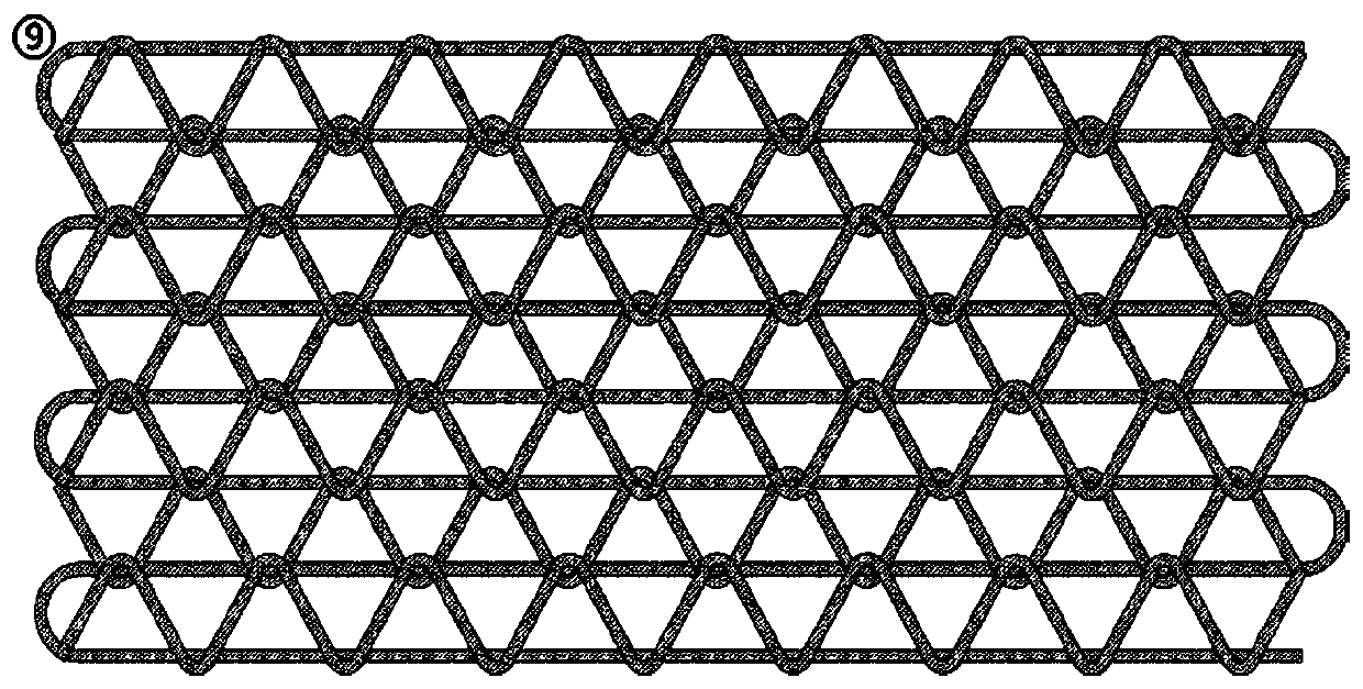
[0045] In this example, if figure 2 , image 3 Among them, the electrospinning described in the step (2) adopts wave-shaped and linear stacked arrangements.

[0046] In this example, if figure 2 , image 3 Among them, the cartilage patch support in step (2) adopts a hollow structure, and the shape of ...

Embodiment 2

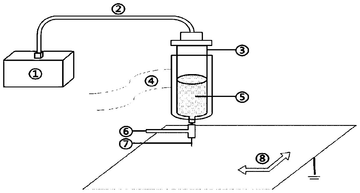
[0052] see figure 1 As shown, this embodiment provides a cartilage patch scaffold construction device based on polycaprolactone electrospinning 3D printing, including a material bucket 3 and a table moving device 8, the material bucket 3; the bottom of the material bucket 3 is provided with a printer nozzle 7 and a high-voltage electrode 6; the high-voltage electrode 6 and the table grounding device on the table moving device 8 jointly build an electric field strength to form electrospinning.

[0053] In this example, if figure 1 In the step (2), the 3D printing device further includes: an air pump device 1, a material barrel 3 connected to the air pump device 1 through an air guide tube 2, and a heating device 4 for heating the material 5 in the material barrel 3 , a printer nozzle 7, a table mobile device 8 and a control unit equipped with 3D printing intelligent control software.

[0054] In this example, if figure 1 Among them, a control unit and a heating device 4 are ...

Embodiment 3

[0060] Using the polycaprolactone electrospinning 3D printing cartilage patch scaffold construction method described in Example 1 to construct a cartilage patch scaffold for clinical use includes the following steps.

[0061] (1) Individually design the shape and structure of the patch as needed, and build a 3D model.

[0062] (2) Through 3D printing combined with electrospinning technology, using PCL as a raw material, the pre-built 3D model was printed to form a cell scaffold.

[0063] (3) Extract primary chondrocytes for expansion, plant the amplified chondrocytes on PCL cell scaffolds, and culture them in a specific environment. Chondrocytes are amplified on the scaffolds and secrete extracellular matrix to form chondrocytes- Extracellular matrix-scaffold complex.

[0064] (4) The complex is transplanted subcutaneously for vascularization, so that it has more perfect biological performance.

[0065] (5) Orthotopic transplantation of the vascularized chondrocyte-extracell...


PUM


Abstract

The invention discloses a method for constructing a cartilage patch stent based on polycaprolactone electrostatic spinning 3D printing and a device thereof. The method comprises the following steps: (1) designing an individualized 3D model of a stent according to characteristic parameters of the stent based on individual characteristics; (2) by utilizing an electrostatic spinning 3D printing technology and taking polycaprolactone as a raw material, printing the cartilage patch stent according to the individualized 3D model; (3) inducing the cartilage cells to be planted on the cartilage patchstent, amplifying the cartilage cells on the stent, secreting extracellular matrixes, and forming a cartilage cell-extracellular matrix-support complex. According to the preparation method, polycaprolactone is taken as the raw material, a technical means of combining electrostatic spinning and 3D printing is employed so as to construct the cartilage patch, and the prepared cartilage patch is widerin application range and excellent in biomechanical property.

Description

technical field [0001] The invention relates to the field of medical technology, in particular to a method and device for constructing a cartilage patch scaffold based on polycaprolactone electrospinning 3D printing. Background technique [0002] Cartilage tissue has few cells, no blood vessels and nerve tissue distribution, and mainly relies on the osmosis of extracellular matrix to provide nutrients. Therefore, it is difficult to regenerate articular cartilage tissue damage caused by acute and chronic injuries or degenerative diseases; in addition, clinically For the repair of long trachea, direct anastomosis cannot be performed, and tracheal cartilage substitutes are also required for repair. At present, the clinical repair of articular cartilage mainly includes: microfracture surgery, osteochondral transplantation and periosteum transplantation, etc., and the repair of tracheal cartilage mainly includes: autologous tissue, allogeneic tracheal transplantation, etc., but t...

Claims


Application Information

Patent Timeline
Patent Type & Authority Applications(China)
IPC IPC(8): A61L27/18A61L27/38A61L27/36A61L27/50B33Y10/00B33Y70/10B33Y80/00D04H1/728D04H1/76D04H1/435
CPCA61L27/18A61L27/3633A61L27/3654A61L27/3817A61L27/3852A61L27/50A61L2430/06A61L2430/40B33Y10/00B33Y70/00B33Y80/00D04H1/435D04H1/728D04H1/76C08L67/04
Inventor 陈昶汤海佘云琅任怡久苏杭孙惟言
Owner SHANGHAI PULMONARY HOSPITAL
Why Eureka
Try Eureka