Novel three-modal prostate cancer targeted nanoparticle developer and preparation method thereof

A nanoparticle, prostate cancer technology, applied in the fields of nuclear medicine, nanomedicine, and radiation medicine, can solve the problems of small molecular weight, difficult long-term continuous imaging, and low tumor uptake

Active Publication Date: 2020-03-24
BEIJING CANCER HOSPITAL PEKING UNIV CANCER HOSPITAL
View PDF4 Cites 5 Cited by
  • Summary
  • Abstract
  • Description
  • Claims
  • Application Information

AI Technical Summary

Problems solved by technology

Commonly used PET and MRI contrast agents have small molecular weights, are cleared quickly in...

Method used


Image

  • Novel three-modal prostate cancer targeted nanoparticle developer and preparation method thereof
  • Novel three-modal prostate cancer targeted nanoparticle developer and preparation method thereof
  • Novel three-modal prostate cancer targeted nanoparticle developer and preparation method thereof

Examples


preparation example Construction

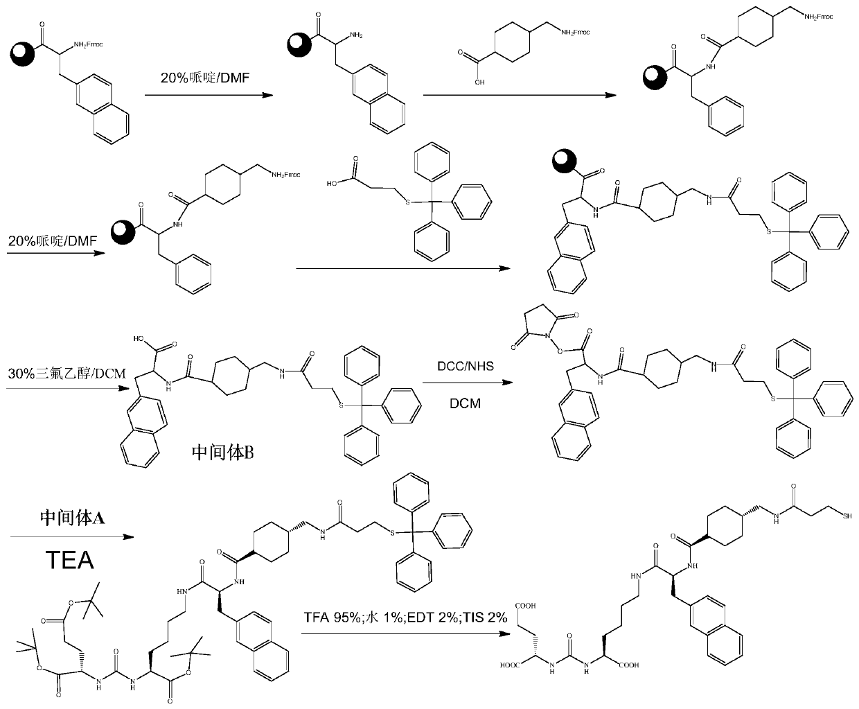
[0064] The present invention also provides a three-modal tumor-targeting nanoparticle imaging agent, which uses the tumor-targeting nanoparticle as a labeling precursor, and Mn 2+ coupling and 89 Zr nuclide labeling. The preparation method comprises the following steps:

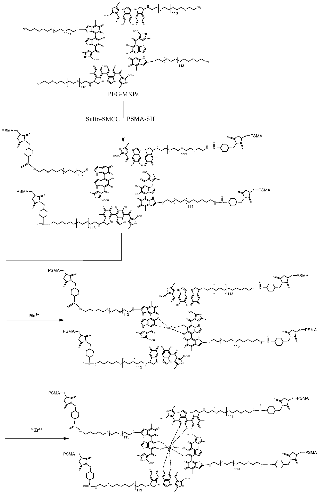
[0065] 1. Targeted modification of nanoparticles: PSMA small molecule inhibitors were modified with sulfhydryl groups to obtain PSMA-SH, and then PSMA-SH was coupled to the surface of SMCC-PEG-UMNPs nanoparticles to form tumor-targeting nanoparticles PSMA-PEG - UMNPs, separated by PD-10 column as labeling precursor.

[0066] 2. Mn 2+Coupling: melanin nanoparticles have a high affinity for metal ions, and can directly conduct electrophilic reactions without coupling agents for Mn 2+ Coupled to obtain Mn-PSMA-PEG-UMNPs nanoprobes with magnetic resonance T1 weighted imaging function.

[0067] 3. 89 Zr labeling: also using the high affinity of melanin nanoparticles to metal ions, directly 89 Zr nuclide lab...

Embodiment 1 3

[0089] Example 1 Preparation method of trimodal prostate cancer targeting nanoparticle imaging agent

[0090] Trimodal prostate cancer targeting nanoparticle imaging agent ( 89 Zr, Mn)-PSMA-PEG-UMNPs preparation comprises the following steps:

[0091] 1. Preparation of PSMA-PEG-UMNPs

[0092] 1.1 Preparation of UMNPs

[0093] Using melanin extracted from plant cells as raw material, ultrafine-sized UMNPs were prepared by ultrasonication. Biological extraction of melanin: Take 10 mg of melanin and dissolve it in 3 mL of NaOH solution (0.1 M) under vigorous stirring. Under the action of an ultrasonic cell pulverizer (working intensity 15%, power 20W), add about 2.5mL of 0.1M HCl solution within 1 minute, adjust the pH of the system to 7.5, and obtain a black and bright UMNPs dispersion; use a molecular weight cut-off of 30kDa The ultrafiltration centrifuge tube removes free Na in the solution + , Cl - , and washed twice with deionized water to obtain pure ultrafine melanin...

Embodiment 2 3

[0130] Example 2 Preparation method of three-modal prostate cancer targeting nanoparticle imaging agent

[0131] The preparation method is the same as in Example 1, only the NH in step 1 2 -The molar ratio of amino group and cross-linking agent Sulfo-SMCC on the surface of PEG-UMNPs was changed to 1:30, the molar ratio of SMCC-PEG-UMNPs nanoparticles to PSMA-SH was changed to 1:30, PSMA-PEG-UMNPs in step 2 Nanoparticles and MnCl 2 The molar ratio was changed to 1:1000, and the remaining reaction conditions were the same.

[0132] The results showed that the number of PSMA-SH coupled to a single nanoparticle increased from 20 to 23, and the Mn 2+ The coupling number did not change significantly. Income ( 89 The labeling rate and radiochemical purity of Zr, Mn)-PSMA-PEG-UMNPs were both greater than 95%.


PUM

PropertyMeasurementUnit
The average particle sizeaaaaaaaaaa

Abstract

The invention provides a novel three-modal prostate cancer targeted nanoparticle developer and a preparation method thereof. With ultra-micro-particle-size organic melanin nanoparticles (UMNPs) with endogenous and high biological compatibility as a carrier, a small molecular group with a PSMA targeting function is coupled with nanoparticles to obtain a nanometer molecular image probe PSMA-PEG-UMNPs with the prostate cancer targeting function; nuclide<89>Zr and T1 weighted magnetic resonance contrast agent Mn<2+> are marked directly by using a UMNPs surface active group to obtain a nanoprobe (<89>Zr, Mn)-PSMA-PEG-UMNPs with PAI, MRI and PET three-mode imaging functions. The probe can be specifically bound with a PSMA antigen on the surface of a prostate cancer cell; the PSMA high-expressiontissues are located accurately by optical, nuclear magnetic and nuclear medicine ways; and thus targeted multi-modal molecular imaging diagnosis of tumors is realized.

Description

technical field [0001] The invention relates to the fields of radiation medicine, nuclear medicine and nanomedicine, in particular to a three-mode prostate cancer targeting nanoparticle imaging agent and a preparation method thereof. Background technique [0002] The research on molecular imaging mostly focuses on optical imaging, nuclide imaging and magnetic resonance imaging. Optical imaging is a new type of non-invasive imaging technology, but it still lacks tissue penetration; nuclide imaging has high sensitivity and plays an important role in whole body imaging, but lacks tissue resolution; MRI has ultra-high tissue resolution. The resolution can clearly show the anatomical structure and boundary of the lesion, but the sensitivity is poor and the ability of molecular imaging is lacking. In the field of multimodal imaging and the diagnosis and treatment of prostate cancer, specific small molecule probes targeting prostate specific membrane antigen (PSMA) have made major...

Claims


Application Information

Patent Timeline
IPC IPC(8): A61K49/00A61K49/18A61K49/22A61K51/06A61K51/08A61K51/12B82Y20/00B82Y40/00
CPCA61K49/0002A61K49/186A61K49/1866B82Y20/00B82Y40/00A61K49/225A61K51/1251A61K51/08A61K51/065
Inventor 杨志夏雷朱华蒋金泉郭晓轶
Owner BEIJING CANCER HOSPITAL PEKING UNIV CANCER HOSPITAL
Why Eureka
Try Eureka