Method and device for efficiently separating nucleus pulposus primary cells

A technology of primary cells and nucleus pulposus, applied in the field of efficient separation of nucleus pulposus primary cells, can solve the problems of damage to the integrity of nucleus pulposus primary cells, difficulty in in vitro culture, cumbersome operation, etc., and achieve good proliferation rate and cell shape, The operation process is simple and fast, and the effect of simple equipment

Active Publication Date: 2020-03-24
SHANDONG MEDICAL BIO TECH RES CENT
View PDF2 Cites 0 Cited by
  • Summary
  • Abstract
  • Description
  • Claims
  • Application Information

AI Technical Summary

Problems solved by technology

Although patients with lumbar disc herniation are relatively common, they are older, the nucleus pulposus tissue in the intervertebral disc is aging and the number of nucleus pulposus cells is small, and it is difficult to culture in vitro
However, the traditional primary culture of nucleus pulposus cells is cumbersome, has a high contamination rate, and is prone to failure.
Moreover, the primary nucleus pulposus cells are generally cultured in vitro after 3-4 passages, and the cell morphology gradually changes, which is not conducive to the needs of scientific research. In order to ensure the reliability of the experimental results, cells that have been passed 1-2 times are usually used. nucleus pulposus
[0005] In the prior art, the method for isolating primary cells of the nucleus pulposus is commonly used to digest with trypsin and / or collagenase. Since the cell membrane itself contains protein, the longer the digestion, the enzyme digestion solution can hydrolyze this type of protein. After the protein is hydrolyzed, the cell membrane of the primary nucleus pulposus cells ruptures, which seriously damages the integrity of the primary nucleus pulposus cells, thereby affecting the viability of the isolated primary nucleus pulposus cells and subsequent proliferation and culture of the primary nucleus pulposus cells; and , as the digestion time prolongs, it will also damage the respiratory enzymes of the cells, thereby affecting the metabolism of the cells

Method used


Image

  • Method and device for efficiently separating nucleus pulposus primary cells
  • Method and device for efficiently separating nucleus pulposus primary cells
  • Method and device for efficiently separating nucleus pulposus primary cells

Examples


Embodiment 1

[0060] Experimental method: Aseptically remove the nucleus pulposus tissue, put it into a sterile petri dish with a diameter of 10cm, wash it with normal saline several times until there is no obvious blood stain, and carefully separate the cartilage and other tissues. Add 1ml of 0.25% trypsin solution to make the content of trypsin 0.2U / mL nucleus pulposus tissue, cut the nucleus pulposus tissue into pieces with a diameter of less than 1mm with sterile surgical scissors for about 20 minutes.

[0061] Use a 3ml sterile pasteurized tube to transfer the shredded nucleus pulposus tissue into a 50ml sterile centrifuge tube, and then add digestion solution to the petri dish, so that elastase 0.5U / mL shredded nucleus pulposus tissue, type II collagenase 0.5U / mL minced nucleus pulposus tissue, hyaluronidase 0.2U / per mL minced nucleus pulposus tissue, β-N-acetylglucosaminidase 0.2U / per mL minced nucleus pulposus tissue, wash The remaining nucleus pulposus tissue in the dish was also t...

Embodiment 2

[0065] Experimental method: The nucleus pulposus tissue was aseptically removed and placed in normal saline, washed several times until there was no obvious blood stains, and cartilage and other tissues were carefully separated. Add 1ml of 0.25% trypsin to make the content of trypsin 0.2U / mL of nucleus pulposus tissue, and use sterile surgical scissors to cut the nucleus pulposus tissue into pieces with a diameter of less than 1mm for about 20 minutes.

[0066] Use a 3ml sterile pasteurized tube to transfer the shredded nucleus pulposus tissue into a 50ml sterile centrifuge tube, and then add digestion solution to the petri dish to make elastase 0.4U / mL of shredded nucleus pulposus tissue and type II collagenase 0.4U / mL minced nucleus pulposus tissue, hyaluronidase 0.15U / per mL minced nucleus pulposus tissue, β-N-acetylglucosaminidase 0.15U / per mL minced nucleus pulposus tissue washing dish The remaining nucleus pulposus tissue was also transferred into a 50ml sterile centrifu...

Embodiment 3

[0070] Experimental method: The nucleus pulposus tissue was aseptically removed and placed in normal saline, washed several times until there was no obvious blood stains, and cartilage and other tissues were carefully separated. Add 1ml of 0.25% trypsin to make the content of trypsin 0.1U / mL of tissue, and use sterile surgical scissors to cut the nucleus pulposus tissue into pieces with a diameter of less than 1mm for about 20 minutes.

[0071]Use a 3ml sterile pasteurized tube to transfer the shredded nucleus pulposus tissue into a 50ml sterile centrifuge tube, and then add digestion solution to the petri dish, so that 0.6U of elastase / mL of the shredded nucleus pulposus tissue and type II collagenase 0.6U / mL minced nucleus pulposus tissue, hyaluronidase 0.3U / per mL minced nucleus pulposus tissue, β-N-acetylglucosaminidase 0.3U / per mL minced nucleus pulposus tissue washing dish The remaining nucleus pulposus tissue was also transferred into a 50ml sterile centrifuge tube with...


PUM


Abstract

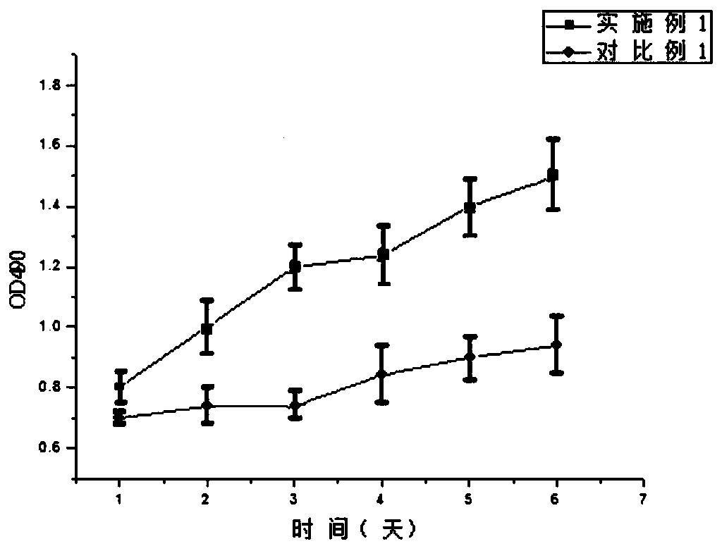
The invention relates to a method for efficiently separating nucleus pulposus primary cells. The method comprises the following steps: a digestion step, wherein active ingredients of a digestive juiceadopted in the digestion step comprise elastase, II-type collagenase and glycosidase; compared with a traditional nucleus pulposus primary cell separation method, the method disclosed by the invention is simple and rapid in operation process, mild in digestion process, simple in required instrument and equipment, and relatively low in cost; the obtained nucleus pulposus cells are large in quantity, high in survival rate, easier to adhere to the wall in a culture bottle, good in proliferation rate and cell morphology and high in cell activity.

Description

technical field [0001] The disclosure relates to a method and a device for preparing primary nucleus pulposus cells, in particular to a method for efficiently separating primary nucleus pulposus cells and a tissue shredding device. Background technique [0002] The information disclosed in this Background section is only intended to increase the understanding of the general background of the disclosure, and is not necessarily to be taken as an acknowledgment or any form of suggestion that the information constitutes prior art that is already known to those skilled in the art. [0003] Low back pain is the most common disease in orthopedics. It not only causes mental pain to patients, but also causes huge economic burden. However, there is still no effective treatment in clinical practice. The intervertebral disc is the largest avascular organ in the human body. Studies have reported that about 40% of patients' low back pain is caused by the degeneration of the intervertebral...

Claims


Application Information

Patent Timeline
IPC IPC(8): C12N5/077C12M1/00
CPCC12N5/0655C12M47/04C12N2509/00C12N2509/10
Inventor 王丹丹曹盛楠师浩钧王从安王磊磊邹亮张波张庆浩任鹏程黄沁周军
Owner SHANDONG MEDICAL BIO TECH RES CENT
Why Eureka
Try Eureka