Method for isolating extracellular vesicles using cations

A technology for separating cells and cations, which is applied in the field of separating extracellular vesicles by using cations, which can solve the problems of difficult and efficient development of antibodies and protein ligands, low efficiency, and high cost

Pending Publication Date: 2020-05-12
ROSETTA EXOSOME CO LTD
View PDF4 Cites 10 Cited by
  • Summary
  • Abstract
  • Description
  • Claims
  • Application Information

AI Technical Summary

Problems solved by technology

Methods for extracellular vesicle isolation using these antibodies or proteins are inefficient, making it difficult and costly to develop antibodies and protein ligands efficiently, so they are very limited

Method used


Image

  • Method for isolating extracellular vesicles using cations
  • Method for isolating extracellular vesicles using cations
  • Method for isolating extracellular vesicles using cations

Examples


Embodiment 1

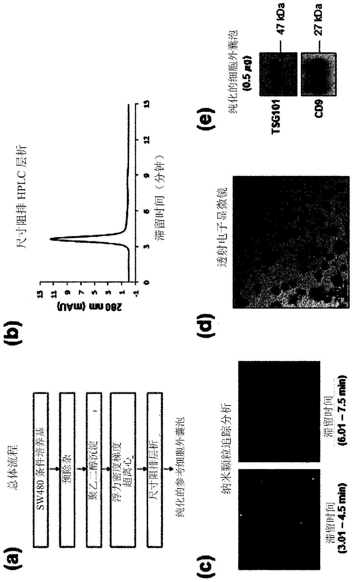
[0069] Example 1: Purification and Analysis of Sample Extracellular Vesicles

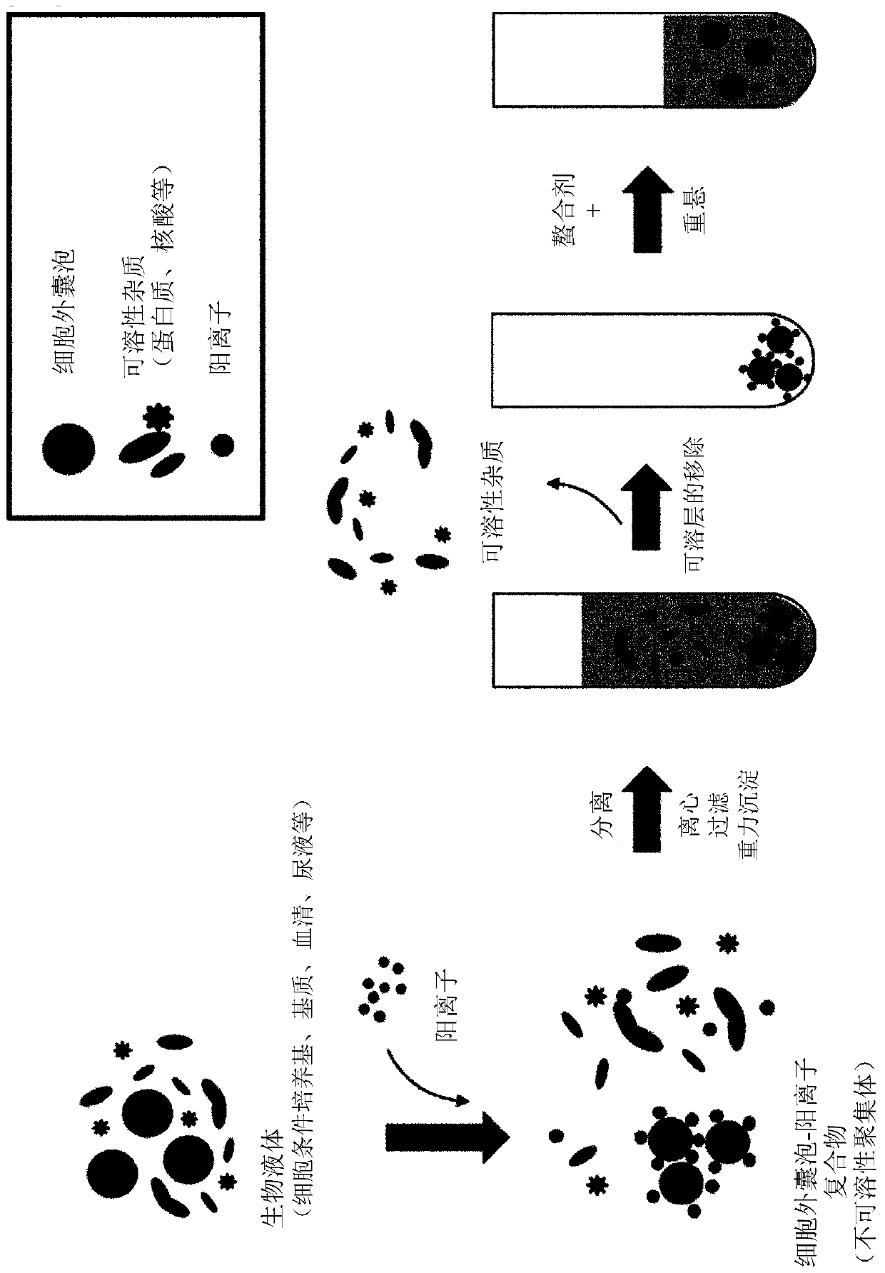
[0070] Colorectal cancer cell SW480 cultures were centrifuged at 500 x g for 10 min (total of two repetitions) to remove residual cells and pellets. The supernatant was centrifuged again at 2,000 xg for 20 minutes (totally repeated twice) to remove the precipitate.

[0071] For the preliminary purification and precipitation of extracellular vesicles present in the supernatant, extracellular vesicle precipitation induction solution (8.4% polyethylene glycol 6000, 250 mM NaCl, 20 mM HEPES, pH 7.4) was added to the supernatant, After 16 hours of storage in the refrigerator, the pelleted extracellular vesicles were collected by centrifugation at 12,000 xg for 30 minutes, and then dissolved in HEPES-buffered saline (20 mM HEPES, 150 mM NaCl, pH 7.4).

[0072] For the second purification of extracellular vesicles using density and buoyancy, the sample obtained in the previous step was mixed with Optiprep...

Embodiment 2

[0076] Example 2: Isolation of extracellular vesicles using several concentrations of different types of cations

[0077] Several concentrations of different types of cations (Ca 2+ 、Cu 2+ , Zn 2+ ), and centrifuged at 3,000×g for 10 minutes to collect the precipitate, which was then dissolved in HEPES buffered saline containing 50 mM EDTA. image 3 A shows the analysis results of extracellular vesicles separated by size exclusion chromatography using the HPLC system, and the extracellular vesicles in the sample were detected at 3.6 minutes.

[0078] Regarding the treatment concentrations of calcium, copper and zinc cations added to colorectal cancer cell cultures, it was confirmed that the 280 nm absorption band detected at 3.6 minutes increases proportionally with the concentration of cations, these results are as follows image 3 B to 3D as shown. The absorption bands of various types of cations and the absorption bands of sample EVs showed the same time of detection, a...

Embodiment 3

[0079] Example 3: Isolation of extracellular vesicles using copper cations (copper(II) chloride)

[0080] Add several concentrations of copper cations to colorectal cancer cell cultures, mix well and centrifuge at 3,000×g for 10 minutes to collect the precipitate, which is then dissolved in HEPES buffered saline containing 50 mM EDTA. Extracellular vesicles isolated by the method described above were studied by nanoparticle tracking analysis and Western blot analysis. Nanoparticle tracking analysis was carried out using NanosightLM10 instrument, and under the conditions of camera level 10 and detection limit 3, tracking and recording were carried out for 60 seconds. For western blot analysis, the signal of CD9, a universal extracellular vesicle label, was analyzed after SDS electrophoresis.

[0081] The result is as Figure 4 As shown in A, the production of extracellular vesicles increases with increasing copper cation concentration. Hence in Figure 4 In B, it is demonst...


PUM


Abstract

The present invention relates to a method for isolating extracellular vesicles using cations, and more particularly, to a method for isolating extracellular vesicles from various samples by using theaffinity between the extracellular vesicles and cations. A method for isolating extracellular vesicles according to the present invention does not require expensive equipment, can be applied irrespective of sample amount, and has the advantage of being capable of efficiently isolating the extracellular vesicles while preserving the shape or characteristics thereof. Moreover, the method according to the present invention can be combined with existing isolation methods to maximize extracellular vesicle isolation efficiency, and can be applied to disease diagnosis, disease treatment, and multi-omics research using isolated extracellular vesicles, as well as to research on the properties of extracellular vesicles.

Description

technical field [0001] The invention relates to a method for separating extracellular vesicles by using cations, in particular to a method for separating extracellular vesicles from various samples by using the affinity of extracellular vesicles to various types of cations. Background technique [0002] Extracellular vesicles are nano-sized vesicles that are naturally released by all living organisms or cells, from humans to bacteria, through pervasive cellular mechanisms. Extracellular vesicles derived from eukaryotic cells are involved in the differentiation of erythrocytes, the regulation of immune responses, etc., especially in the microenvironment of cancer cells, which play an important role in the development, metastasis or angiogenesis of cancer. Therefore, the application of extracellular vesicles as diagnostic labels in various diseases including cancer has received much attention. [0003] Similar to extracellular vesicles of eukaryotic cells, extracellular vesic...

Claims


Application Information

Patent Timeline
Patent Type & Authority Applications(China)
IPC IPC(8): C12N5/00B01D15/36
CPCB01D15/362B01J39/00C12N5/00B01D15/36C12N2509/00G01N1/40
Inventor 高用松李昌津金志贤宋成炫
Owner ROSETTA EXOSOME CO LTD
Why Eureka
Try Eureka