
Method for nondestructive analysis of mesenchymal stem cell membrane protein

A cell membrane and protein technology, applied in the field of cell detection, can solve problems affecting membrane proteins and cell activity, and achieve the effect of convenient experimental operation.

Active Publication Date: 2020-06-05
章毅 +7
View PDF4 Cites 0 Cited by
  • Summary
  • Abstract
  • Description
  • Claims
  • Application Information

AI Technical Summary

Problems solved by technology

But this method requires irreversible labeling of the target protein with an antibody, which can unpredictably affect the activity of membrane proteins and cells

Method used


Image

  • Method for nondestructive analysis of mesenchymal stem cell membrane protein
  • Method for nondestructive analysis of mesenchymal stem cell membrane protein
  • Method for nondestructive analysis of mesenchymal stem cell membrane protein

Examples


Embodiment 1

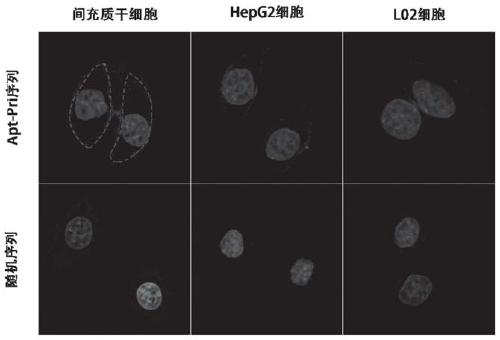
[0054] Example 1 Qualitative detection of mesenchymal stem cell membrane protein

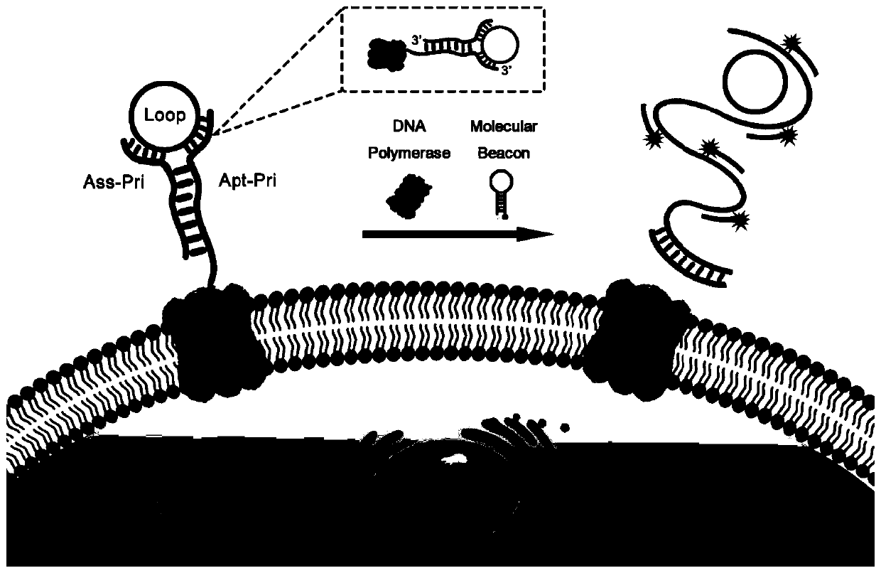
[0055] The aptamer primer (Apt-Pri) was heated at 95°C for 5 minutes to denature it, and then cooled to room temperature to form a DNA secondary structure. In glass-bottom confocal dishes according to 1 × 10 per dish 5 cells were inoculated. In order to allow the cells to adhere to the wall better, they need to be cultured in a carbon dioxide incubator at 37°C for 24 hours. To prevent the endocytosis of aptamers, 150 μL of 100 μM chlorpromazine hydrochloride was added to the medium and incubated with the cells for 30 min in a CO2 incubator.

[0056] Wash twice with 1 × PBS to remove the medium containing chlorpromazine hydrochloride, and then add 150 μL of aptamer-protein binding buffer (containing 1 mg / mL bovine serum albumin (BSA) containing 150 nM aptamer primer (Apt-Pri) , 10v / v% fetal bovine serum (FBS), 4.5g / L glucose and 5mM magnesium chloride (MgCl 2 ) serum-free high-glucose medium ...

Embodiment 2

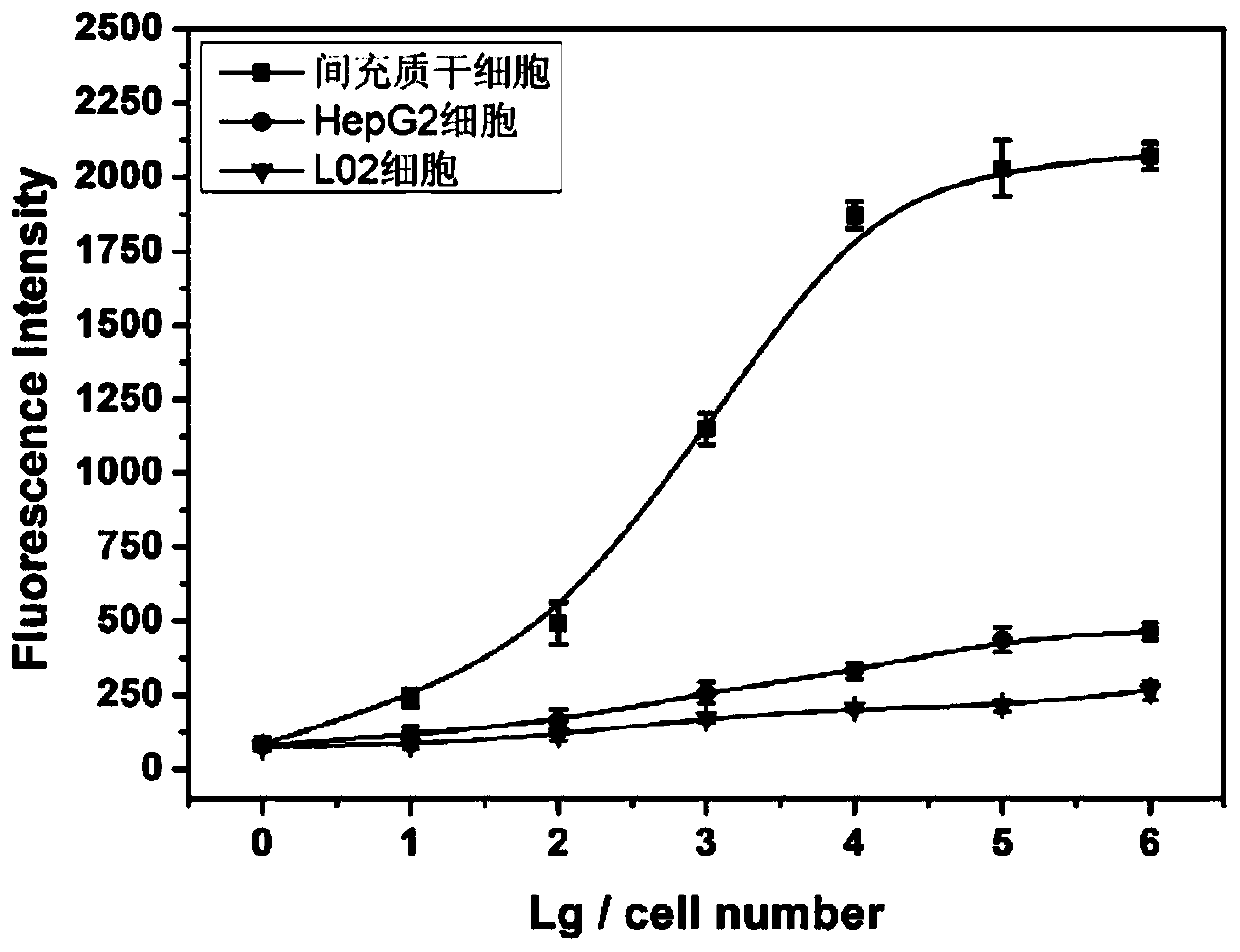
[0057] Example 2 Quantitative detection of mesenchymal stem cells

Embodiment 3

[0060] Example 3 Nondestructive Analysis of Mesenchymal Stem Cell Membrane Proteins

[0061] Firstly, three groups of mesenchymal stem cells, HepG2 and L02 cells were prepared, and the CD44 protein of the three kinds of cells in the first group was determined by Western Blot experiment. Then, the first round of analysis was performed on the three types of cells in the second group according to the method in the above-mentioned Example 2, and the cells after the analysis were determined by Western blotting. Then carry out the first round of analysis on the three types of cells in the second group according to the means in the above-mentioned Example 2. After the analysis, the cells are placed in a carbon dioxide constant temperature cell incubator to continue culturing for 2 hours, and continue to follow the above example after the state is stable. The method in 2 was used for the second round of analysis, and the cells after the analysis were determined by Western blotting. T...


PUM


Abstract

The invention discloses a method for analyzing cell membrane protein. The cell membrane protein comprises an aptamer primer as shown in SEQ ID No 1, an auxiliary primer as shown in SEQ ID No 2, a circular DNA template as shown in SEQ ID No 3 and a molecular beacon as shown in SEQ ID No 4. According to the method, the membrane protein of the cell is recognized through a DNA chain, and an amplifiedfluorescence signal is obtained, so that nondestructive analysis of the target cell membrane protein is realized, the method can be customized according to specific detection requirements of target cells (such as mesenchymal stem cells and even membrane proteins on the surfaces of other different types of cells), actual requirements of different analysis are met, and the method has good application potential.

Description

technical field [0001] The invention relates to a method for detecting cells, in particular to a method for analyzing cell membrane proteins, so as to realize the non-destructive analysis of mesenchymal stem cells. Background technique [0002] Mesenchymal stem cells (MSCs) originate from the stem cells of the embryonic mesoderm and exist widely in the body. Among them, mesenchymal stem cells belong to a type of pluripotent stem cells of the mesoderm, mainly exist in connective tissue and organ interstitium, concentrate in bone marrow and adipose tissue, and have multiple potentials. Under specific conditions, it can be induced to differentiate into different types of cells in vivo and in vitro, such as: chondrocytes, osteoblasts, adipocytes, nerve cells and cardiomyocytes. It is easy to isolate and culture, and has a high potential for in vitro expansion. Clinical experiments have confirmed that mesenchymal stem cell transplantation can be used for tissue repair and treat...

Claims


Application Information

Patent Timeline
IPC IPC(8): G01N21/64G01N15/10G01N33/50G01N33/68
CPCG01N21/6458G01N15/10G01N33/5091G01N33/6842G01N2015/1006Y02A50/30
Inventor 章毅朱小立伍婷任灵杰陈侃俊刘晓浩陈亮
Owner 章毅
Learn More