Liquid sample cell for microscopic Raman spectroscopy detection of aerobic or facultative anaerobic pathogenic microorganisms

A technology of pathogenic microorganisms and liquid samples, which is applied in the field of liquid sample pools, to achieve the effects of reducing environmental pollution, having biological safety, and slowing down sample volatilization

Inactive Publication Date: 2021-05-04
INST OF MICROBIOLOGY - CHINESE ACAD OF SCI
View PDF11 Cites 0 Cited by
  • Summary
  • Abstract
  • Description
  • Claims
  • Application Information

AI Technical Summary

Problems solved by technology

[0004] The purpose of the present invention is to provide a liquid sample pool for microscopic Raman spectroscopy detection of aerobic or facultative anaerobic pathogenic microorganisms in view of the problems existing in the sample support vessel in the existing microscopic Raman cell detection

Method used


Image

  • Liquid sample cell for microscopic Raman spectroscopy detection of aerobic or facultative anaerobic pathogenic microorganisms
  • Liquid sample cell for microscopic Raman spectroscopy detection of aerobic or facultative anaerobic pathogenic microorganisms
  • Liquid sample cell for microscopic Raman spectroscopy detection of aerobic or facultative anaerobic pathogenic microorganisms

Examples


Embodiment 1

[0045] Embodiment 1 liquid sample cell

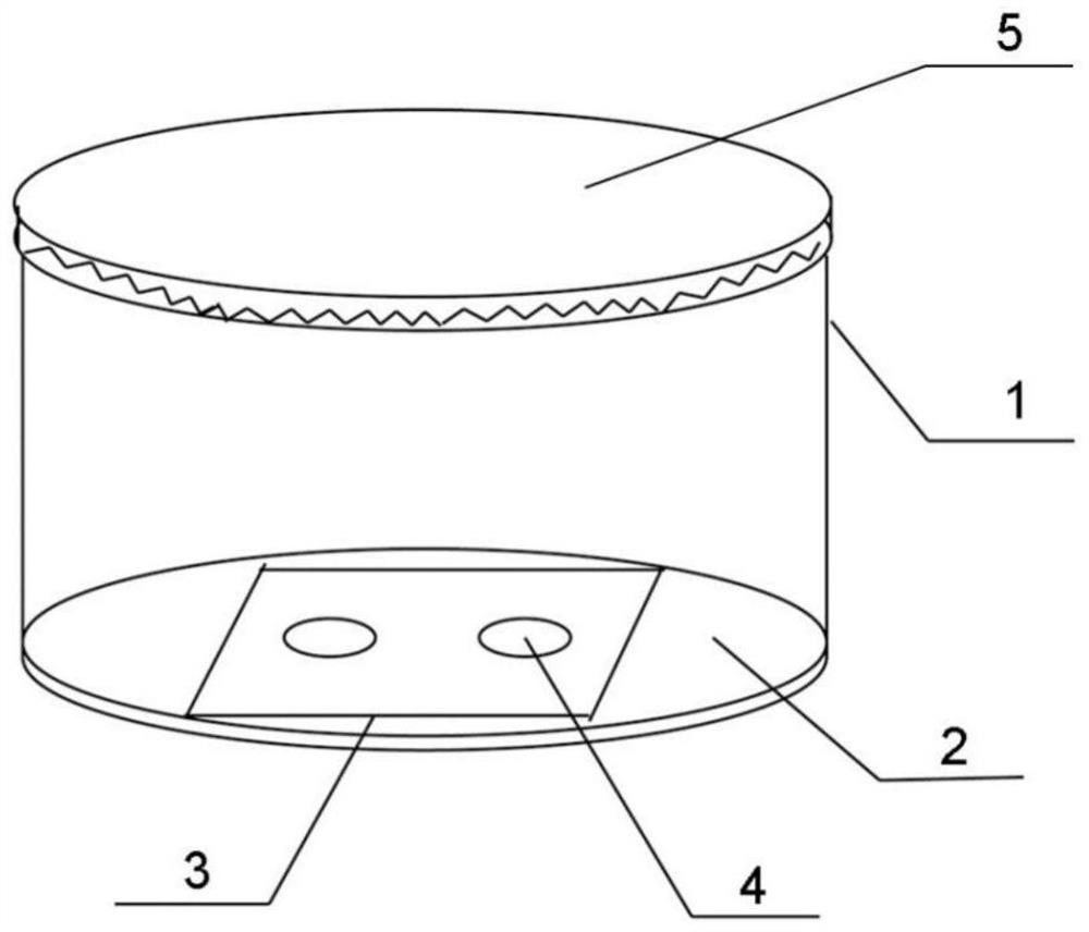
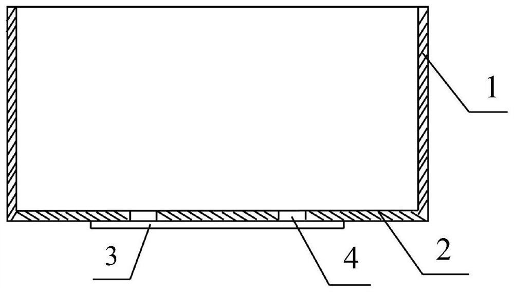
[0046] Such as figure 1 and figure 2 As shown, the liquid sample cell is composed of a sample cell body, a top cover 5 and a double-sided polished cover glass 3 . A quartz slide is selected as the cover glass 3, the sample cell body and the top cover 5 are both formed of polystyrene, and the top cover 5 is connected to the top of the sample cell body in a rotating and sealed manner. The diameter of the sample cell body is 30 mm, and the height is 15 mm. The sample cell body has a side wall 1 and a bottom wall 2, and the thickness of the bottom wall 2 is 1.5 mm. The bottom wall 2 is provided with two circular through-holes 4 with a diameter of 3mm, and the cover glass 3 is adhered to the outer surface of the bottom wall 2 with epoxy acrylate UV glue and covers the through-holes 4, thereby forming two Detection hole. Wherein, the thickness of the cover glass 3 is 0.13 mm, the length is 22.5 mm, and the width is 4.5 mm.

Embodiment 2

[0047] The property test of embodiment 2 liquid sample cell

[0048] Add the concentration of 20 μl to a detection hole of the liquid sample pool of embodiment 1 and be the red pigment water droplet of 3 μ g / ml, screw on the top cover (recorded as " test 1 "); Add 20 μl of red pigment water droplets with a concentration of 3 μg / ml into the sample pool (with no detection hole), and cover the top cover (the top cover has no screw-on structure and cannot be screwed tightly) as a control (referred to as "control"). A micro sampler (with a range of 10 μl) was used to measure the amount of sample remaining in the liquid sample pool, and the results are shown in Table 1.

[0049] Table 1 Sample retention under different storage times

[0050] initial sample size 1 hour reserve 2 hours reserve 3 hours retention test 1 20μl 17.8μl 15.8μl 12.5μl control 20μl 12.3μl 5.5μl 0μl

[0051] It can be known from Table 1 that the liquid sample ...

Embodiment 3

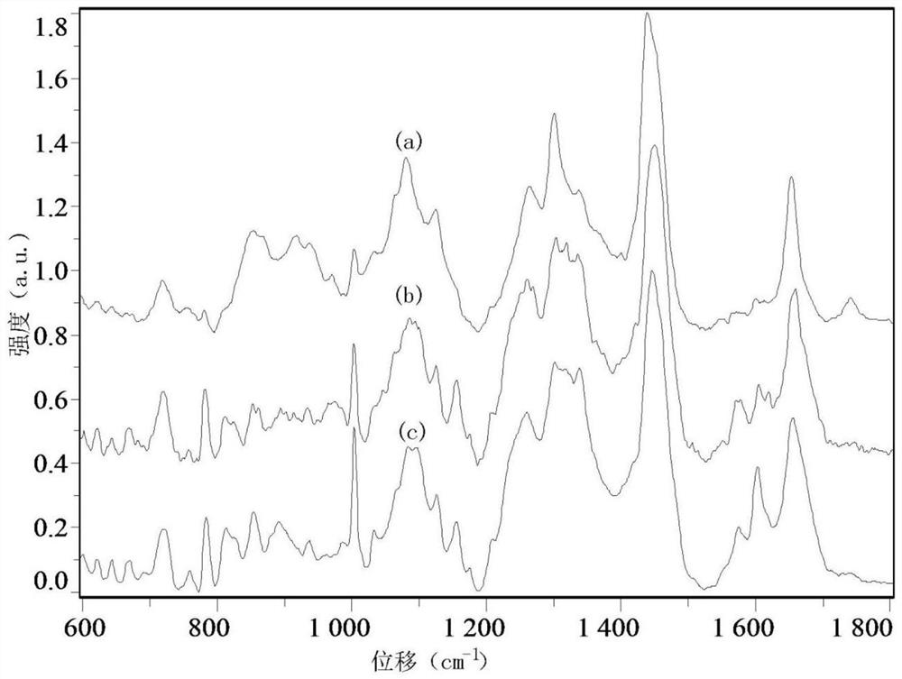
[0052] Example 3 Collection of Cell Raman Spectrum

[0053] (1) Wash one or more samples of pathogenic bacteria once with physiological saline, and suspend the pathogenic bacteria in the physiological saline.

[0054] (2) Add 20 μl of the sample with pathogenic bacteria into the detection hole of the liquid sample pool;

[0055] (3) The liquid sample pool is placed on a micro-Raman spectrometer to capture a single cell to obtain a Raman spectrum.

[0056] Figure 3-4 The Raman spectra of different fungi and bacteria measured by the above method are shown, among which (a) is the Raman spectrum of Cryptococcus neoformans, (b) is the Raman spectrum of Candida albicans, (c) is the Raman spectrum of S. The Raman spectrum of yeast, (d) is the Raman spectrum of Acinetobacter baumannii, and (e) is the Raman spectrum of Pseudomonas aeruginosa.

[0057] Depend on Figure 3-4 It can be seen that the liquid sample pool of the present invention can be used to detect microscopic Raman...


PUM

PropertyMeasurementUnit
widthaaaaaaaaaa
widthaaaaaaaaaa
diameteraaaaaaaaaa

Abstract

The invention provides a liquid sample pool for microscopic Raman spectrum detection of aerobic or facultative anaerobic pathogenic microorganisms, the liquid sample pool includes a sample pool body and a cover glass, and the cover glass is a quartz glass slide Or a calcium fluoride glass slide, the sample cell body has side walls and a bottom wall, the bottom wall is provided with one or more through holes, the cover glass is adhered to the outer surface of the bottom wall and The through holes are covered to form one or more detection holes. The liquid sample pool of the invention has low cost, simple operation, can be used for single-sample or multi-sample measurement, and has biological safety.

Description

technical field [0001] The invention belongs to the field of Raman spectrum analysis, in particular to a liquid sample pool used for micro-Raman spectrum detection of aerobic or facultative anaerobic pathogenic microorganisms. Background technique [0002] Raman spectroscopy is developed based on the principle of inelastic scattering caused by inelastic collisions between photons and chemical molecules. Through the microscope, microscopic samples can be observed under the microscopic structure, such as cells, protozoa, microorganisms, etc. After the laser is irradiated on the microscopic samples, the samples can be excited to produce Raman scattering, and the scattered light forms bands after being separated. Mann spectroscopy can determine the vibrational modes of molecules, and then infer the structural information of molecules. Micro-Raman spectroscopy has the advantages of fast, simple, repeatable and non-destructive analysis, and requires a small amount of sample (for ...

Claims


Application Information

Patent Timeline
Patent Type & Authority Patents(China)
IPC IPC(8): G01N21/65
CPCG01N21/65G01N2021/651
Inventor 付钰李焓菲卢维来
Owner INST OF MICROBIOLOGY - CHINESE ACAD OF SCI
Why Eureka
Try Eureka