Method of detecting membrane protein of extracellular vesicles

A membrane protein and cell technology, which can be used in measurement devices, biological tests, material inspection products, etc.

Inactive Publication Date: 2020-06-23
北京益微生物科技有限公司
View PDF1 Cites 10 Cited by
  • Summary
  • Abstract
  • Description
  • Claims
  • Application Information

AI Technical Summary

Problems solved by technology

The above-mentioned traditional EVs isolation and protein detection methods have their limitations: EVs isolation requires a large sample size, cumbersome operation, and time-consuming; and it is difficult to achieve high-throughput detection of EVs membrane proteins
For example, Western blotting can only detect one membrane protein in one experiment, which has the disadvantages of cumbersome operation, low detection throughput, and low sensitivity.
Protein mass spectrometry requires high protein purity, while the traditional separation method of EVs often introduces foreign protein contamination

Method used


Image

  • Method of detecting membrane protein of extracellular vesicles
  • Method of detecting membrane protein of extracellular vesicles
  • Method of detecting membrane protein of extracellular vesicles

Examples


Embodiment 1

[0086] The preparation of embodiment 1 antibody chip

[0087] The positive control biotin-labeled IgG antibody concentration was 0.04 μg / ml, 0.2 μg / ml, 1 μg / ml, 5 μg / ml, and the negative control was PBS solution. The specific antibody concentration against EVs membrane protein was 200μg / ml, dissolved in PBS containing 5% glycerol. The manufacturer of the specific antibody is Sino Biological Inc. The details of the name and article number of the specific antibody are shown in Table 1.

[0088] Table 1 Name and catalog number of specific antibody

[0089]

[0090]

[0091]

[0092]

[0093]

[0094]

[0095]

[0096]

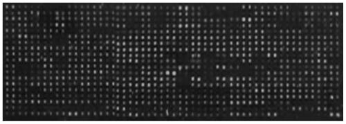
[0097] The solid phase carrier adopts NEXTERION epoxy-coated glass slides from SCHOTT Company of Germany. The above-mentioned antibodies are spotted on the coated glass slides according to a certain arrangement using an automatic spotting instrument, and two replicates are set for each antibody spot.

Embodiment 2

[0098] Embodiment 2 sample pretreatment

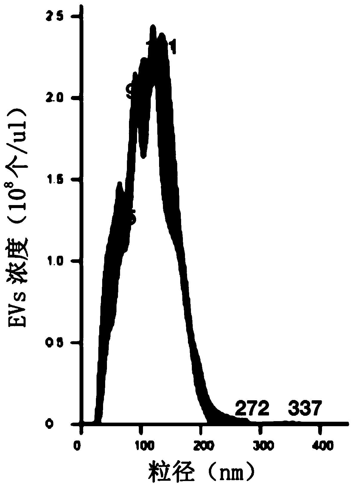
[0099]This example is for the preliminary separation of EVs from the CCC-HEL-1 cell culture supernatant: collect the CCC-HEL-1 cell culture supernatant, centrifuge at 300g for 10min, collect the supernatant, and remove the cells; then centrifuge at 3000g for 10min, and collect the supernatant , remove cell debris; then centrifuge at 10000g for 30min, collect supernatant to remove subcellular components; then concentrate with 100KD ultrafiltration centrifuge tube, wash with PBS 3 times, and concentrate to about 100 times in final volume.

[0100] Then, the particle size and concentration analysis of the EVs obtained from the preliminary separation was carried out using the Malvern nanoparticle tracking analyzer NanoSight LM14. Test results such as figure 1 As shown, the concentration of EVs in the pretreated samples was 2.3×10 8 particles / μl, the particle size is mainly distributed around 150nm.

Embodiment 3

[0101] Example 3 Antibody Chip Separation of EVs and Analysis of EVs Membrane Protein Composition

[0102] Add the pre-treated samples to the antibody chip to isolate EVs. The specific method is as follows:

[0103] (1) Adding samples: the concentration of pretreated EVs added to the antibody chip was 10 5 -10 8 per μl, the volume added was 100 μl. Incubate at room temperature for 2 h on a horizontal shaker at a low speed (60 rpm / min). Then transfer to 4°C and continue to incubate for 18h.

[0104] (2) Washing: Aspirate the sample from the sample pool, add 100 μl of PBS solution containing 0.2% Tween20 to wash 3 times, 5 min each time.

[0105] (3) Detection: Add a mixture of detection antibodies (biotin-labeled antibodies against EVs membrane protein markers CD9, CD63, and CD81), and incubate at room temperature for 2 hours on a horizontal shaker at a low speed (60 rpm / min). Aspirate the biotin-labeled antibody mixture from the sample pool, and repeat the washing proces...


PUM


Abstract

The invention relates to the field of biological detection, in particular to a method for detecting membrane protein of extracellular vesicles. The method comprises the following steps: a) co-incubating a composition containing extracellular vesicles (EVs) with a solid phase carrier; wherein the solid phase carrier is coated with a first antibody group of anti-EVs membrane protein; b) washing offnon-specifically bound EVs and other impurities, and c) adding a second antibody group for detection to carry out signal detection, the second antibody group being an antibody of a biomarker of EVs and coupled with a marker for displaying signal intensity. The method is simple and convenient to operate, the detection flux can be flexibly adjusted according to conditions, and the sensitivity is high.

Description

technical field [0001] The invention relates to the field of biological detection, in particular to a method for detecting the membrane protein of extracellular vesicles. Background technique [0002] Extracellular vesicles (EVs) are membrane vesicle structures of 20-1000nm in size secreted by cells, and are widely distributed in cell culture supernatants and various body fluids (blood, urine, saliva, etc.) . EVs are mainly composed of proteins, nucleic acids, and phospholipid molecules, and serve as transmitters of genetic material and information between cells [Colombo M, Raposo G, Théry, Clotilde. Biogenesis, Secretion, and Intercellular Interactions of Exosomes and Other Extracellular Vesicles [J]. Annual Review of Cell and Developmental Biology, 2014, 30(1):255-289.]. [0003] EVs play important biological functions and participate in the regulation of various biological processes and disease processes, such as tumors, neurological diseases, cardiovascular diseases, a...

Claims


Application Information

Patent Timeline
Patent Type & Authority Applications(China)
IPC IPC(8): G01N33/68G01N33/58G01N33/532
CPCG01N33/532G01N33/58G01N33/6842
Inventor 樊萌
Owner 北京益微生物科技有限公司
Why Eureka
Try Eureka