A kind of extracellular vesicle derived from human amniotic mesenchymal stem cells and its application
A technology of stem cells and human amniotic membrane, applied in the field of biomedicine to achieve the effect of inhibiting the hypertrophy of cardiomyocytes
- Summary
- Abstract
- Description
- Claims
- Application Information
AI Technical Summary
Problems solved by technology
Method used
Image
Examples
Embodiment 1
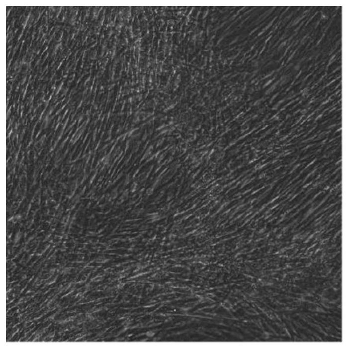
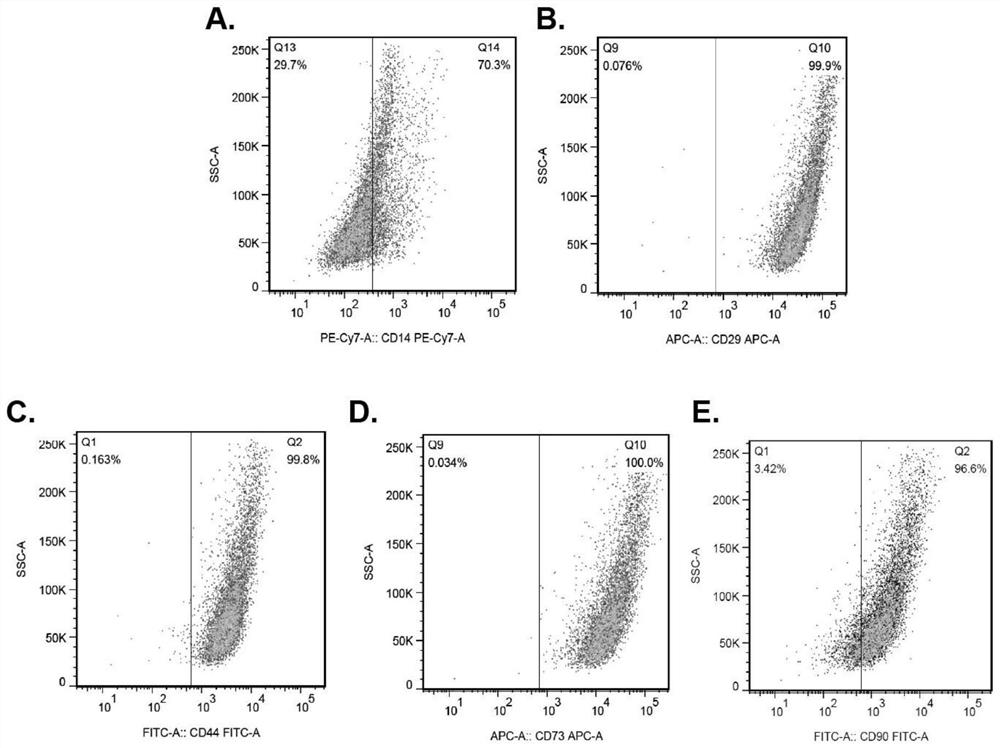
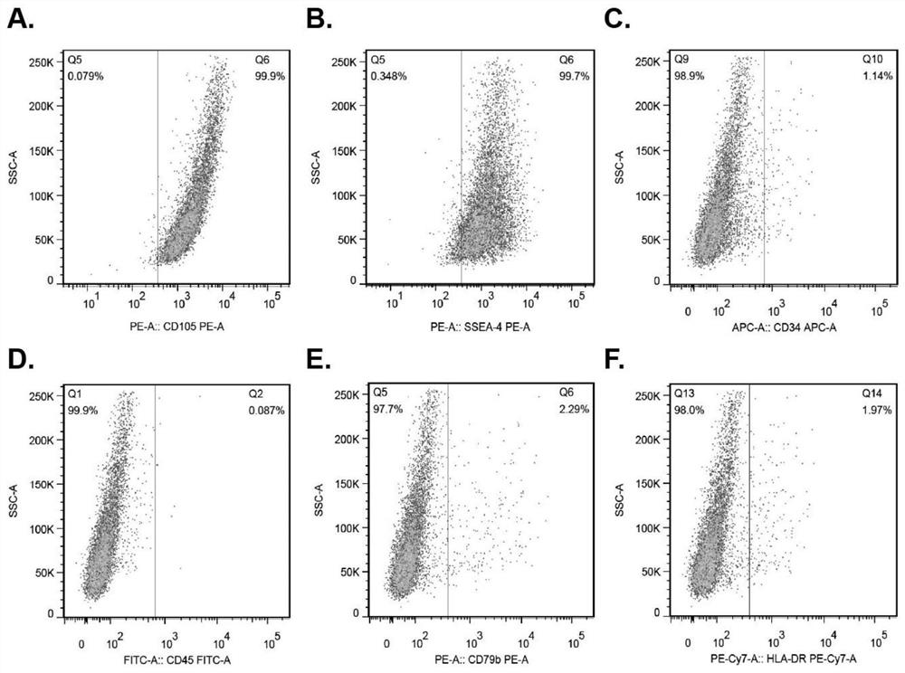
[0045] Example 1 Preparation of Hamscs-EVS of Human Ferrous Metachate Stem Cell Source
[0046] Separation of Hamscs:
[0047] (1) Rinse the amniotic tissue repeatedly, then cut the amniotic tissue, then cut the amniotic membrane, according to the plasma of the amniotic membrane: (1-2), the volume ratio of the digestion, 37 ° C Incubate 7-10 min, obtained after digestion; the digestion is a PBS containing 2.4 U / mL protease DISPase;
[0048] (2) Add a digestion of amniotic tissue to α-MEM complete medium containing 10% fetal bovine serum, after standing for 5-10 min, according to amniotic tissue: Digestion = 1: (2-3) volume Digestion was digested at 37 ° C to give 0.75 mg / mL collagenase COLLAGEN D and 5% fetal bovine serum α-MEM complete medium;
[0049] (3) Cut the tissue liquid after digestion (1500 rpm is centrifuged for 10min), go to the supernatant, leave the precipitate, and repeatedly rinsed, centrifuge, precipitate until it is clear, abandon the supernatant, with 15% fe...
Embodiment 2
[0063] Example 2Hamscs-EVS is transmitted into a hypertrophy of intraocular intraocular cells via cyclone
[0064] Hamscs-EVS, DII, using fluorescently active dyes DII (1'-Dioctadecyl-3, 3, 3 ', 3'-Tetramethylindocarbocyanine Perchlorate), DII is one of the most commonly used cell membrane fluorescent probes, presenting orange-red fluorescent, entering cell membrane After late diffusion, the cell membrane of the entire cell can be gradually dyed, and the extracted Hamscs-EVS extracted in Example 1 was resuspended with PBS, and 1 μM DII dye was 37 ° C, including 5% CO. 2In the cell incubator, it was incubated for 5 min and then incubated into centrifuge muscle, centrifuged at 4 ° C, 100000 g from 1 h, abandoned out excess of dye, washed with PBS 3 times, and the PBS was resuspended. Dii marked Hamscs-EVS co-cultured with myocardial hypertrophy of cells, PBS was rinsed 3 times, and the cell membrane of myocardial fertilizer (WGA) was labeled with WGA staining with WGA, and laser Foc...
Embodiment 3
[0066] Example 3Hamscs-EVS prevention and pathological myocardial cell hypertrophy
[0067] Construct a cardiac hypertrophy: In order to clearly construct a highly treated concentration concentration of cardiac III, use 5 × 10 -7 M, 5 × 10 -6 M and 1 × 10 -5 M Vastrial Angiothen II (Ang II) The three gradient concentrations induce human myocardial cell AC16 hypertrophy, and the compass of myocardial fertilizer major cell model was determined by Real-Time PCR. Figure 8 After Hamscs-EVS, the cardiocyte membrane was labeled by wheat nodule (WGA), and the laser confocal microscope was observed in the cell surface area, and the results showed that 5 × 10 -6 Mang II Treats AC16 Heartocytes, with 5 × 10 -7 M and 1 × 10 -5 Compared to cell hypertrophic phenotype, the cell surface area has increased significantly ( Figure 8 . Figure 9 Cardiomyopathic peptide (ANP), brain sodium urine peptide (BNP), β-myocardin heavy chain (BNP), β-myocardium (β-MHC) and myocardium β-MHC MRNA and myocardial...
PUM
| Property | Measurement | Unit |
|---|---|---|
| diameter | aaaaa | aaaaa |
| diameter | aaaaa | aaaaa |
Abstract
Description
Claims
Application Information
 - R&D
- Intellectual Property
- Life Sciences
- Materials
- Tech Scout
- Unparalleled Data Quality
- Higher Quality Content
- 60% Fewer Hallucinations
Browse by: Latest US Patents, China's latest patents, Technical Efficacy Thesaurus, Application Domain, Technology Topic, Popular Technical Reports.
© 2025 PatSnap. All rights reserved.Legal|Privacy policy|Modern Slavery Act Transparency Statement|Sitemap|About US| Contact US: help@patsnap.com


