Axitinib-contained nanofiber electrospun membrane and preparation method and application thereof

A nanofiber and axitinib technology, which is applied in the field of nanofiber electrospun membrane containing axitinib and its preparation, can solve the problems of easy ossification of cartilage and achieve ossification prevention, stable structure and simple operation Effect

Inactive Publication Date: 2020-08-21
SHANGHAI CHILDRENS MEDICAL CENT AFFILIATED TO SHANGHAI JIAOTONG UNIV SCHOOL OF MEDICINE
View PDF7 Cites 2 Cited by
  • Summary
  • Abstract
  • Description
  • Claims
  • Application Information

AI Technical Summary

Problems solved by technology

[0007] In order to solve the problem of easy ossification of cartilage regeneration in the subcutaneous environment, the present invention provides a nanofibrous material that is biocompatible, degradable and absorbable, and can significantly inhibit angiogenesis

Method used


Image

  • Axitinib-contained nanofiber electrospun membrane and preparation method and application thereof
  • Axitinib-contained nanofiber electrospun membrane and preparation method and application thereof
  • Axitinib-contained nanofiber electrospun membrane and preparation method and application thereof

Examples


Embodiment 1

[0039] Embodiment 1: Preparation of collagen / polycaprolactone nanofiber membrane loaded with Axitinib

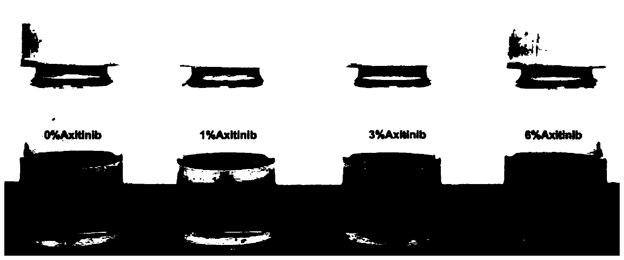
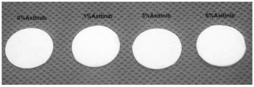
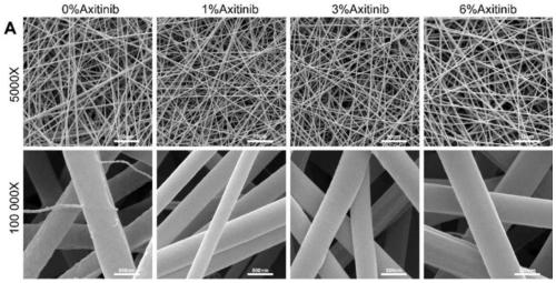
[0040] 0 mg, 12 mg, 36 mg, and 72 mg of Axitinib were weighed and dissolved in 10 ml of hexafluoroisopropanol, and 20-30 microliters of acetic acid was added to the solution, and stirred until uniformly mixed. Then add 0.48 gram of collagen and 0.72 gram of polycaprolactone to be made into the spinning solution (collagen and polycaprolactone total mass and hexafluoroethylene) of 12% g / ml in the Axitinib solution of above-mentioned four kinds of different concentrations to be made into mass volume percentage then respectively volume percent of isopropanol solution). A 10ml syringe and a needle with an inner diameter of 1.2mm were selected to extract the collagen / polycaprolactone spinning solution, which was fixed on an electrospinning device for spinning. Electrospinning parameters are: voltage 8-15kv, receiving distance 10-15cm, injection rate 1-3ml / h, temperature 15-30°C, ...

Embodiment 2

[0042] Example 2: Study on in vitro biocompatibility and in vitro activity of collagen / polycaprolactone nanofiber membrane loaded with Axitinib

[0043] Rabbit bone marrow mesenchymal stem cells (BMSCs) and rabbit auricle chondrocytes were isolated, cultured and expanded in vitro. The collagen / polycaprolactone nanofiber membranes loaded with 0%, 1%, 3% and 6% Axitinib were sterilized and placed at the bottom of a 24-well plate for use. The second-generation BMSCs and second-generation chondrocytes were seeded on four different amounts of Axitinib-loaded nanofiber materials. On the 1st, 4th and 7th day after inoculation, add CCK8 solution to the culture medium for incubation, and detect the absorbance of the supernatant to judge the proliferation efficiency of the cells on the material. On day 1 and day 7 after inoculation, the cells were fixed, and the nuclei and cytoskeleton were fluorescently stained and photographed.

[0044] The result is as Figure 4 As shown, compared...

Embodiment 3

[0047] Example 3: Construction of tissue engineered cartilage blocks derived from stem cells in vitro

[0048] Use cylindrical PGLA non-woven scaffolds, such as Figure 6 As shown in A. Observation of the scaffold with a scanning electron microscope, such as Figure 6 B-C, it can be seen that the fiber thickness is uniform, and the fiber diameter distribution is measured, such as Figure 6 D, PGLA fiber diameter is 12.7±1.8um. The second-generation BMSCs were seeded on PGLA scaffolds, such as Figure 6 E, in vitro chondrogenic induction culture, at intervals, SEM observation of cell attachment growth on the scaffold, such as Figure 6 F-H( Figure 6 F is the first day of cell inoculation, Figure 6 G is the 7th day of inoculation, Figure 6 H is the 14th day of inoculation). It can be seen that with the increase of culture time, the secretion of cell matrix increases and gradually fills the gaps between PGLA fibers. After 4 weeks of chondrogenic induction, the tissue-...


PUM


Abstract

The invention provides an axitinib-contained nanofiber electrospun membrane. The nanofiber electrospun membrane consists of axitinib, collagen and polycaprolactone. The invention further provides a preparation method and application of the nanofiber electrospun membrane. The nanofiber electrospun membrane has good biocompatibility, can be degraded and absorbed and can remarkably inhibit blood vessel generation, and therefore the problem that cartilages are prone to ossification when regenerated in a subcutaneous environment is solved.

Description

technical field [0001] The invention relates to the technical field of biomedical materials, in particular to a nanofiber electrospun membrane containing axitinib and its preparation method and application. Background technique [0002] Cartilage is an avascular, anervous, and alymphoid tissue with cells tightly embedded in a highly dense natural extracellular matrix (ECM). Due to the poor regeneration ability of cartilage, its repair and reconstruction has always been a major problem in clinical treatment. Tissue engineering technology provides a new way for the repair of cartilage defects, among which articular cartilage repair has initially achieved clinical transformation, but there has been no breakthrough for cartilage defects in the subcutaneous environment (such as cartilage defects in ears, nose, trachea, etc.) progress. The main dilemma of subcutaneous cartilage regeneration is that when the cartilage tissue constructed from stem cells with clinical application p...

Claims


Application Information

Patent Timeline
Patent Type & Authority Applications(China)
IPC IPC(8): D04H1/4382D04H1/728A61L27/18A61L27/22A61L27/54D01F1/10
CPCA61L27/18A61L27/222A61L27/54A61L2300/216A61L2300/602A61L2430/06D01F1/10D01F6/92D04H1/4382D04H1/728C08L89/00C08L67/04
Inventor 李奋冯蓓冀天基朱荻绮陈轶维吉炜张浩
Owner SHANGHAI CHILDRENS MEDICAL CENT AFFILIATED TO SHANGHAI JIAOTONG UNIV SCHOOL OF MEDICINE
Why Eureka
Try Eureka