Bionic exosome as well as preparation and application thereof

An exosome and bionic technology, applied in the field of bionic exosome, can solve problems such as undiscovered tumor treatment, achieve the effect of increasing tumor self-targeting ability, avoiding phagocytosis, and increasing circulation performance

Active Publication Date: 2021-02-26
THE SECOND XIANGYA HOSPITAL OF CENT SOUTH UNIV
View PDF3 Cites 4 Cited by
  • Summary
  • Abstract
  • Description
  • Claims
  • Application Information

AI Technical Summary

Problems solved by technology

At present, there are no reports on this type of biomimetic exosomes, l

Method used


Image

  • Bionic exosome as well as preparation and application thereof
  • Bionic exosome as well as preparation and application thereof
  • Bionic exosome as well as preparation and application thereof

Examples


Embodiment 1

[0060] This example is used to illustrate the preparation of liposomes.

[0061] Liposomes were prepared by the thin film dispersion method. 20mg lipid material DPPC, cholesterol and DSPE-PEG 2000 (mass ratio 80:15:5) was dissolved in 2 mL of chloroform:methanol (3:1 V / V), and the solvent was evaporated by a rotary evaporator to form a lipid film.

[0062] Add 2mL PBS to the lipid film for hydration to form a lipid suspension.

[0063] The lipid suspension was passed through a cellulose acetate membrane with a pore size of 100 nm through a liposome extruder at 25°C for 20 times, and the obtained homogeneous liposome solution was stored at 4°C for use.

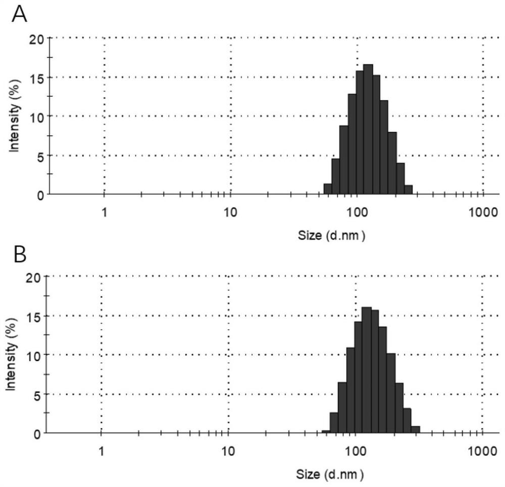
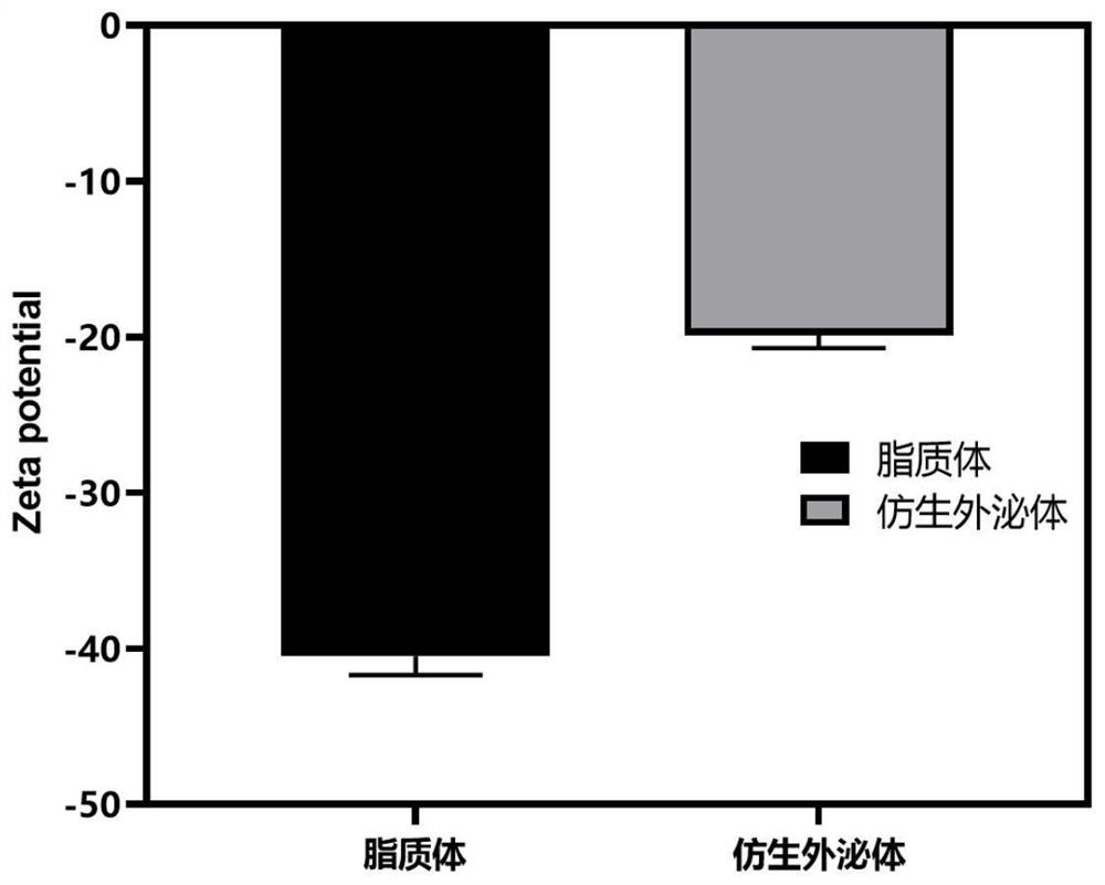
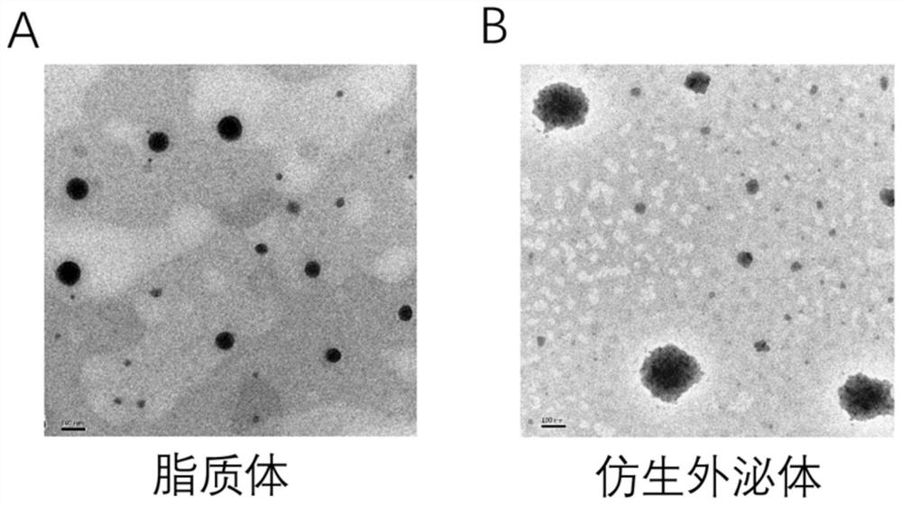
[0064] The particle size distribution of liposomes ( figure 1 A), zeta potential ( figure 2 ) was characterized by a nanometer particle size analyzer, and the morphology ( image 3 A) Characterized by transmission electron microscopy.

[0065] In the particle size distribution diagram, the average particle size of liposo...

Embodiment 2

[0067] This example is used to illustrate the preparation of biomimetic exosomes.

[0068] 4T1 breast tumor cell membrane protein was extracted by cell membrane protein and cytoplasmic protein extraction kit, and the concentration of cell membrane protein was determined by BCA method.

[0069] In Example 1, tumor cell membrane protein was added while PBS was added to the lipid film for hydration, the mass ratio of membrane protein to lipid material was 1:100, and the mixture was fully mixed to form a suspension.

[0070] Add NE to the membrane protein-lipid suspension, the mass ratio of NE to lipid material is 1:40, incubate at 25°C for 30min, then pass through a cellulose acetate membrane with a pore size of 100nm through a liposome extruder for 20 times , and the obtained homogeneous biomimetic exosome solution was stored at 4°C for future use.

[0071] Particle size distribution of biomimetic exosomes ( figure 1 B), Zeta potential ( figure 2 ) was characterized by a nan...

Embodiment 3

[0074] This example is used to illustrate the protein characterization of membrane protein chimeric liposomes in the biomimetic exosomes of the present invention.

[0075] The tumor cell membrane proteins on the biomimetic exosomes prepared in Example 2 of the present invention were determined by Coomassie brilliant blue staining. Configure 10% SDS polyacrylamide gel, load 10 μg of 4T1 breast tumor cell total protein, 4T1 breast tumor cell membrane protein, 4T1 breast tumor cell cytoplasmic protein and biomimetic exosomes, and infiltrate with Coomassie brilliant blue staining solution after electrophoresis Soak and stain for 2 hours, then decolorize with decolorizing solution. Take pictures to observe the protein bands between groups (results see Figure 4 ). The results showed that the protein of the biomimetic exosome was very similar to the membrane protein of 4T1 breast tumor cells, but significantly different from the cytoplasmic protein of 4T1 breast tumor cells, indic...


PUM


Abstract

The invention discloses a bionic exosome as well as preparation and application thereof. An artificial lipid material is subjected to tumor cell membrane protein chimerism to prepare a nano-liposome with a particle size similar to that of an exosome structure, and NE is combined on the surface of the nano-liposome through electrostatic adsorption so as to be used as the bionic exosome. The bionicexosome has a stable lipid membrane structure, tumor targeting and a tumor matrix degradation function. After the bionic exosome degrades a tumor matrix in a targeting manner, the permeation of an anti-cancer drug into a tumor and the immersion of immune cells into the tumor can be enhanced, the anti-tumor curative effect is greatly improved, and the bionic exosome has huge conversion value and application prospect in the aspect of combined chemotherapy/immune anti-tumor.

Description

technical field [0001] The invention belongs to the technical field of biomedicine, and specifically relates to a bionic exosome targeting to degrade tumor matrix and its preparation and application. Background technique [0002] Tumor is a major disease that seriously endangers human health. Desmoplastic tumors have a dense tumor stroma mainly composed of abundant tumor extracellular matrix and tumor-associated fibroblasts, which encloses relatively few tumor cells, and blood vessels are buried in the tumor stroma, not directly adjacent to tumor cells , not only makes it difficult for drugs to penetrate into the tumor, but also hinders the infiltration of immune cells in the tumor site. Using nanotechnology to break through the tumor matrix can promote the entry of anticancer drugs into the tumor, and at the same time promote the infiltration of immune cells in the tumor, which is of great significance for tumor treatment. [0003] Neutrophils are important immune cells i...

Claims


Application Information

Patent Timeline
IPC IPC(8): A61K9/127A61K47/24A61K47/42A61K38/48A61P35/00
CPCA61K9/1271A61K47/24A61K47/42A61K38/486C12Y304/21037A61P35/00
Inventor 向大雄李泳江吴军勇胡雄彬
Owner THE SECOND XIANGYA HOSPITAL OF CENT SOUTH UNIV
Why Eureka
Try Eureka