Shrimp immunofluorescence detection test strip and application

A technology for immunofluorescence detection and test strips, which can be used in biological tests, measuring devices, material inspection products, etc., and can solve problems such as low sensitivity

Pending Publication Date: 2021-04-02
杭州福德敏生物技术有限公司
View PDF0 Cites 1 Cited by
  • Summary
  • Abstract
  • Description
  • Claims
  • Application Information

AI Technical Summary

Problems solved by technology

However, the current allergen detection method can only detect one kind at a time, and the sensitivity is low

Method used


Image

  • Shrimp immunofluorescence detection test strip and application
  • Shrimp immunofluorescence detection test strip and application
  • Shrimp immunofluorescence detection test strip and application

Examples


preparation example Construction

[0022] The preparation method of the conjugate pad of the present invention preferably comprises:

[0023] (a) Adsorbing and combining fluorescently labeled streptavidin and latex microspheres to prepare fluorescent latex microspheres;

[0024] (b) combining biotin with the mixed antibody to prepare a biotinylated mixed antibody;

[0025] (c) mixing the fluorescent latex microspheres with the biotinylated mixed antibody to obtain the mixed antibody labeled with fluorescent latex microspheres;

[0026] (d) Spraying the mixed antibody labeled with fluorescent latex microspheres on the binding pad; there is no time sequence between steps (a) and (b).

[0027] In the present invention, the mass ratio of fluorescently labeled streptavidin and latex microspheres described in step (a) is preferably 1:40; the volume ratio of biotin and mixed antibody described in step (b) is preferably 1:4; the volume ratio of fluorescent latex microspheres and biotinylated mixed antibodies in step ...

Embodiment 1

[0036] 1. Preparation of binding pads

[0037]1) Preparation of fluorescent latex microspheres: Dilute latex microspheres with a particle size of 400 nm to a final concentration of 30 mg / ml with an adsorption buffer (50 mM, citrate buffer at pH 5.8) and a volume of 6 ml to obtain latex microspheres Suspension; add red fluorescein rhodamine-labeled liantavidin in the adsorption buffer according to the volume ratio of 1:50~500, the final volume is 6ml; The mixed solution was prepared in the adsorption buffer solution of mycoavidin labeled clearly; the resulting mixed solution was incubated at room temperature for 1 to 2 hours, and stirred continuously, and then centrifuged to collect the precipitate, and the precipitate was stored in buffer solution (containing 0.06% BSA Adsorption buffer) was dissolved, stored at 4°C, and set aside.

[0038] 2) Preparation of biotinylated mixed antibody

[0039] The antigen myosin antibody (US Indoor, product number: PA-SHM); anti-arginine ki...


PUM

PropertyMeasurementUnit
particle diameteraaaaaaaaaa

Abstract

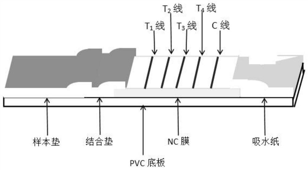
The invention provides a shrimp immunofluorescence detection test strip and application, and relates to the technical field of detection test strips. The immunofluorescence detection test strip comprises a sample pad, a conjugate pad, a nitrocellulose membrane and absorbent paper which are sequentially assembled and adhered to a PVC bottom plate, wherein the conjugate pad is coated with a fluorescent latex microsphere labeled mixed antibody, the nitrocellulose membrane is sequentially coated with an antigen myosin antibody (T1 line), an anti-arginine kinase antibody (T2 line), an anti-hemocyanin antibody (T3 line), an anti-troponin / myosin light chain antibody (T4 line) and a rabbit anti-mouse IgG antibody (C line), the T1 line, T2 line, T3 line and T4 line are quality control lines, and the line C is a quality control line. The test strip can accurately and quantitatively detect the content of tropomyosin, arginine kinase, hemocyanin and troponin / tropomyosin light chains in food, and is simple and convenient to operate, high in accuracy and high in sensitivity.

Description

technical field [0001] The invention belongs to the technical field of detection test strips, in particular to a shrimp immunofluorescence detection test strip and its application. Background technique [0002] The safety of food allergens has attracted widespread attention, and its potential allergenicity is a new topic that urgently needs to be studied. In the past 20 years, the incidence of allergies has increased significantly, and food allergens have become one of the hidden dangers of food safety. With the development of the economy and the improvement of people's living standards, people's attention and attention to allergies has greatly increased. Shrimp, crab and other crustaceans and their products are an important category of the 8 categories of food allergies proposed by the Food and Agriculture Organization of the United Nations. Its allergen is mainly a kind of tropomyosin (Pen al) present in muscle fibers. Food allergy endangers the health of allergic people...

Claims


Application Information

Patent Timeline
Patent Type & Authority Applications(China)
IPC IPC(8): G01N33/573G01N33/68G01N33/543G01N33/558G01N33/533
CPCG01N33/533G01N33/54313G01N33/558G01N33/573G01N33/68G01N2333/4712
Inventor 李欣武涌孟轩夷佟平袁娟丽
Owner 杭州福德敏生物技术有限公司
Why Eureka
Try Eureka