MiRNA detection method based on a solid nanopore sensor, detection probe and kit.

A technology for nanopore sensors and detection probes, which is applied in the directions of DNA/RNA fragments, recombinant DNA technology, microbial measurement/inspection, etc. It can solve the problems of difficult detection, unfavorable high-throughput detection, and high cost of nanopores Improving the effectiveness of early diagnosis and treatment

Pending Publication Date: 2021-05-14
NANJING UNIV OF POSTS & TELECOMM
View PDF12 Cites 3 Cited by
  • Summary
  • Abstract
  • Description
  • Claims
  • Application Information

AI Technical Summary

Problems solved by technology

However, the detection of miRNAs in solid-state nanopores is still difficult. Due to factors such as processing technology and membrane thickness, the background noise of solid-state nanopore electrical signals is large, and the resolution of small molecules such as short DNA sequences is low.
At the same time, when short DNA sequences pass through the solid nanopore, the speed is too fast, and the signal acquisition also has higher requirements
In this regard, people have made a series of improvements to solid-state nanopore chips, such as surface modification, etc., but the cost of modified nanopores is high, and the detection molecule is single, which is not conducive to high-throughput detection.

Method used


Image

  • MiRNA detection method based on a solid nanopore sensor, detection probe and kit.
  • MiRNA detection method based on a solid nanopore sensor, detection probe and kit.
  • MiRNA detection method based on a solid nanopore sensor, detection probe and kit.

Examples


Embodiment 1

[0060] Embodiment 1: the preparation of gold nanoparticle probe and the concrete implementation to miRNA target molecular detection comprise the following steps:

[0061] Step 1: BSPP (3 mg) was added to 10 ml of AuNPs solution (the diameter of AuNPs is 5 nm), and the mixture was shaken overnight at room temperature.

[0062] Step 2: Slowly add sodium chloride to this mixture while stirring until the color changes from dark burgundy to light purple.

[0063] Step 3: The resulting mixture was centrifuged for 5 min (8000 rpm), and then the supernatant was removed. Then add 0.5ml BSPP (2.5mM / L), and add 0.5ml methanol. The mixture was centrifuged again, the supernatant was removed, and 100 uL (2.5 mM / L) of BSPP was added.

[0064] Step 4: Add the DNA fragment shown in SEQ ID NO.1 to BSPP-AuNPs for single-molecule labeling of DNA probes.

[0065] Specifically: (1) First, incubate BSPP-AuNPs with the DNA fragment shown in SEQ ID NO.1 at a molar ratio of 1:10 for 16 hours, and sh...

Embodiment 2

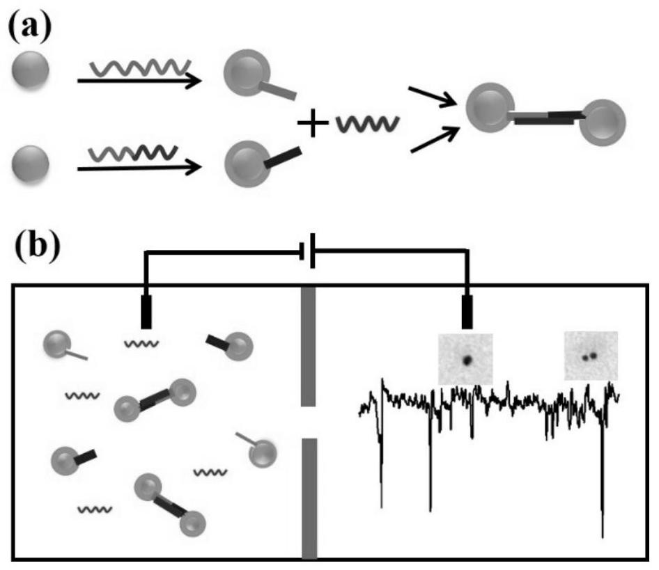
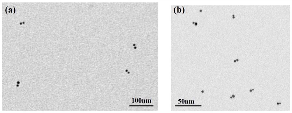
[0077] Example 2: Solid state nanopore detection of miR-21 and miR-486

[0078] (1) miRNA target molecules including miR-21 and miR-486 were added in equal proportions to the designed probe solution, and the mixture was left overnight.

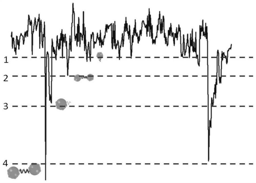
[0079] (2) Add the reacted solution to a silicon nitride solid-state nanopore sensor with a pore size of 40nm, apply different bias voltages of 0-1000mV for signal detection, drive the gold ball through the hole, observe the translocation process of the nanoparticle, and use a diaphragm Clamp records trace current signals. Its sampling frequency is 100KHz, and the cutoff frequency of low-pass filter is 10KHz.

[0080] figure 2 The TEM image of the nanoparticle dimer formed after the gold nanoparticles regulated by the DNA probe bind to the miRNA target molecules, from figure 2 It can be seen from the results that the detection probe designed by the present invention has a strong adsorption capacity on the adenine A sequence at one end of ...


PUM

PropertyMeasurementUnit
particle diameteraaaaaaaaaa
pore sizeaaaaaaaaaa

Abstract

The invention discloses a miRNA detection method based on a solid nanopore sensor, a detection probe and a kit. The detection method provided by the invention comprises the following steps: taking gold nanoparticles as a carrier, and carrying out DNA probe monomolecular marking on different gold nanoparticles. When the probe and the target miRNA coexist, the DNA probe and the miRNA target molecule are hybridized and complemented, and the probe is induced to form a gold nanoparticle dimer. When the gold nanoparticle monomer and the gold nanoparticle dimer pass through the nanopore sensor, different current track characteristic signals are generated, so that the miRNA target molecule is identified. The method can be used for multi-element detection of multiple lung cancer marker miRNA biological target molecules at the same time. According to the invention, the problem that a solid-state nanopore sensor cannot detect miRNA small biomolecules is solved, multi-element joint detection of multiple lung cancer marker miRNA target molecules is realized, and the application of the silicon nitride solid-state nanopore in the field of single molecule detection is expanded.

Description

technical field [0001] The invention relates to a solid-state nanopore sensor detection method, in particular to a solid-state nanopore sensor-based miRNA detection method, a detection probe and a kit. Background technique [0002] miRNAs are short non-protein-coding RNAs that regulate mRNA expression at the transcriptional or post-transcriptional level. Numerous studies have shown that miRNA plays an important role in cell proliferation, differentiation, apoptosis and metabolism. Abnormally expressed miRNAs have been detected in a variety of human malignant tumors, and are related to the occurrence, development, prediction, diagnosis, treatment and postoperative of tumors. Therefore, miRNAs, as potential markers for cancer diagnosis, prediction and treatment, have broad prospects for clinical application. miRNA plays multiple roles in non-small cell lung cancer, including playing the role of tumor-promoting gene or tumor suppressor gene in the process of lung cancer, regu...

Claims


Application Information

Patent Timeline
Patent Type & Authority Applications(China)
IPC IPC(8): C12Q1/6825C12N15/11
CPCC12Q1/6825C12Q2563/137C12Q2525/207C12Q2525/173C12Q2565/607C12Q2565/631
Inventor 翁丽星武灵芝严馨曾祥杰
Owner NANJING UNIV OF POSTS & TELECOMM
Why Eureka
Try Eureka