Multi-sinus combined cerebral venous thrombosis animal model and construction method thereof
An animal model, cerebral vein technology, applied in the field of medicine, to achieve the effect of reducing mortality, simple operation and good stability
- Summary
- Abstract
- Description
- Claims
- Application Information
AI Technical Summary
Problems solved by technology
Method used
Image
Examples
Embodiment 1
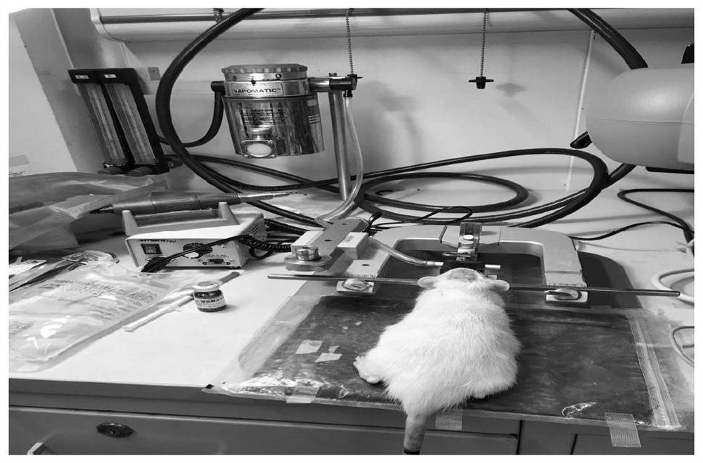
[0051] 1. Enflurane was used to induce anesthesia, and 70% nitrous oxide and 30% oxygen were sent through an anesthesia mask to maintain the entire operation and monitoring process. The anesthetized rats were fixed in a prone position on a stereotaxic frame, and the body temperature was maintained at 37°C with a constant temperature blanket throughout the operation ( figure 1 ).
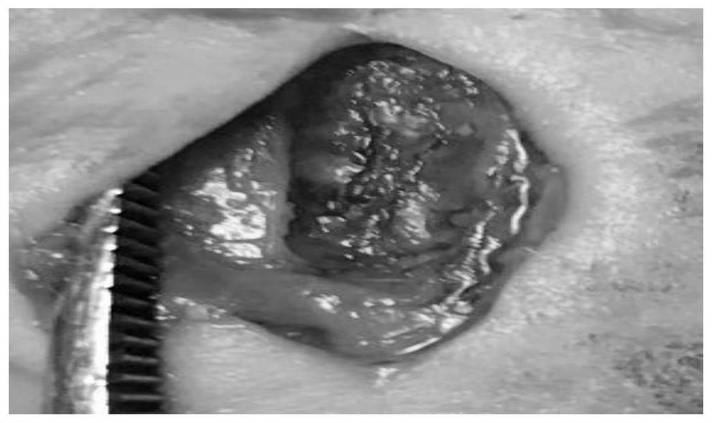
[0052] 2. After skin preparation, make a surgical incision about 1.5cm in the middle of the head, separate the subcutaneous tissue, expose the skull, drill a hole with a grinding drill on the side between the anterior bregma and the herringbone point, operate under a microscope, and gradually expand In order to form a 10mm×3mm bone window, expose the sss and the cortex on both sides, and keep the dura mater intact, the drill bit was continuously cooled with normal saline during drilling to avoid burning the dura mater and cortex ( figure 2 ).
Embodiment 2
[0062] In order to illustrate the repeatability of the animal model construction method of the present invention, table 2 has listed the cerebral blood flow change situation of 20 rats before half ligation, after half ligation and application ferric chloride thrombin, we will half ligation, before half ligation, The cerebral blood flow after semi-ligation and application of ferric chloride thrombin is expressed in the form of mean ± standard deviation, that is, 359.42±72.61PU, 189.98±31.12PU, 95.93±28.31PU, calculated by one-way analysis of variance F =151.90, P Figure 11 ), suggesting that thrombus was formed, indicating that the construction method has good repeatability.
[0063] Table 2. Changes in cerebral blood flow at different stages during modeling
[0064]
Embodiment 3
[0066] For the 20 rat cerebral venous thrombosis animal models constructed in Example 2, 24 hours after modeling, one died of epidural hematoma, and the 24-hour survival rate after operation was 95%. Two each died of severe cerebral ischemia, and the mortality rate at 1 week after operation was 25% (5 / 20), among which the mortality rate due to severe cerebral ischemia was 21.1% (4 / 19), which is similar to that of human severe cerebral ischemia. CVT mortality rates are relatively similar. Twelve hours after the operation, 5 rats were deeply anesthetized and perfused with saline heart. It was found that all 5 rats had thrombosis in the superior sagittal sinus, transverse sinus and internal jugular vein, and even cortical vein thrombosis (see Figure 12 A, The yellow arrow indicates the superior sagittal sinus thrombosis, the green clipped head indicates the transverse sinus thrombosis, the blue arrow indicates the internal jugular vein thrombosis, and the black arrow indicates t...
PUM
 Abstract
Description
Claims
Application Information
 - R&D
- Intellectual Property
- Life Sciences
- Materials
- Tech Scout
- Unparalleled Data Quality
- Higher Quality Content
- 60% Fewer Hallucinations
Browse by: Latest US Patents, China's latest patents, Technical Efficacy Thesaurus, Application Domain, Technology Topic, Popular Technical Reports.
© 2025 PatSnap. All rights reserved.Legal|Privacy policy|Modern Slavery Act Transparency Statement|Sitemap|About US| Contact US: help@patsnap.com


