Tumor combined treatment preparation with cell exosome loaded excitation reagent and photosensitizer and application thereof
A combined therapy and exosome technology, applied in the field of medical biology, to achieve the effect of avoiding infection and small drug resistance
- Summary
- Abstract
- Description
- Claims
- Application Information
AI Technical Summary
Problems solved by technology
Method used
Image
Examples
preparation example Construction
[0036] In step (1), CCExosA is used as an example for the preparation of chemical dynamics and hypoxia linkage treatment preparation, and the preparation method is as follows:
[0037] 1) Separation and extraction of exosomes from M1 microglial cells. Mouse microglial cells BV2 were selected as the research object for culture, and a certain concentration of LPS was added to stimulate the production of M1 microglial cells. No exosomes were used in the whole process. The serum of exosomes was used for cell culture, and exosomes were extracted by ultracentrifugation. The method was as follows: Collect 40 mL of stimulated cell culture supernatant, 300 g, centrifuge at 4°C for 15 min, remove impurities and take the supernatant; 3 000 g, 4 Centrifuge at ℃ for 15 min, remove the cell debris and take the supernatant; centrifuge at 20 000g, 4℃ for 70 min, take the supernatant and filter it with a 0.22 μm sieve; Blot dry, exosomes contained in the pellet, resuspended with appropriate am...
Embodiment 1
[0043] Example 1. Separation and extraction of exosomes
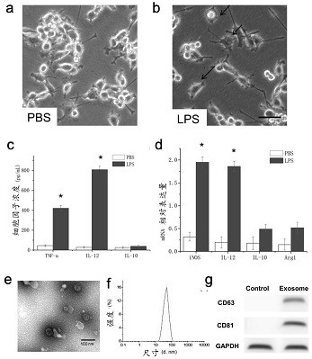
[0044] After LPS (lipopolysaccharide) induction, the morphology of microglial cells changed significantly, showing an amoeba-like activated state, with enlarged cell bodies, retracted processes, and significant increases in pro-inflammatory factors such as TNF-α and IL-12, which manifested as M1 type microglia, and successfully isolated exosomes secreted by M1 type microglia. The toxicity of AQ4N to cells was explored, and it was found that 1 mg / mL of AQ4N had no obvious cytotoxicity after co-incubating with normal microglial cells for 48 h. figure 1 a PBS-treated BV2 was detected for microglial phenotype; figure 1 b is microglial phenotype detection of BV2 induced by LPS; figure 1 c is ELISA detection of TNF-α, IL-12, IL-10 polarizing cytokines; detection of M1 microglial cells; figure 1 d is the expression of related factors detected by RT-PCR, wherein iNOS and Arg1 are M1 markers; figure 1 e is the transmission e...
Embodiment 2
[0045] Example 2. In vivo application of M1-type Exosome regulation of tumor immune microenvironment and chemokinetic therapy preparation
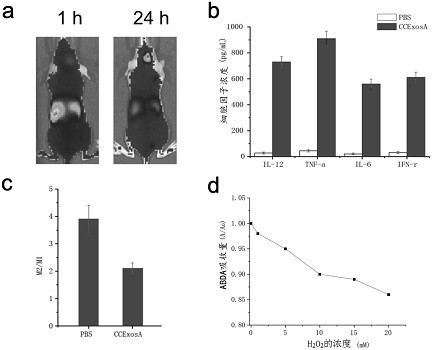
[0046] Tumor-bearing mice were constructed, and CCExosA was injected into the tumor-bearing mice through the tail vein. CCExosA could pass through the BBB to reach the tumor site and be enriched in the tumor site. figure 2 a is the distribution of CCExosA across the BBB and in vivo, and the distribution in vivo over time; figure 2 b is the effect diagram of CCExosA increasing the expression of pro-inflammatory factors; figure 2 c is the change of M2 / M1 ratio in the tumor microenvironment, where M2 is M2 type microglia, M1 is M1 type microglia, and the M2 / M1 ratio in the PBS control group is higher than that of CCExosA, indicating that CCExosA can improve tumor microglia. The secretion of IL-12, IL-6, TNF-α and IFN-γ in the environment reduces the ratio of M2 microglia to M1 microglia in the tumor site, and CCExosA has the function of r...
PUM
 Abstract
Description
Claims
Application Information
 - R&D
- Intellectual Property
- Life Sciences
- Materials
- Tech Scout
- Unparalleled Data Quality
- Higher Quality Content
- 60% Fewer Hallucinations
Browse by: Latest US Patents, China's latest patents, Technical Efficacy Thesaurus, Application Domain, Technology Topic, Popular Technical Reports.
© 2025 PatSnap. All rights reserved.Legal|Privacy policy|Modern Slavery Act Transparency Statement|Sitemap|About US| Contact US: help@patsnap.com


