

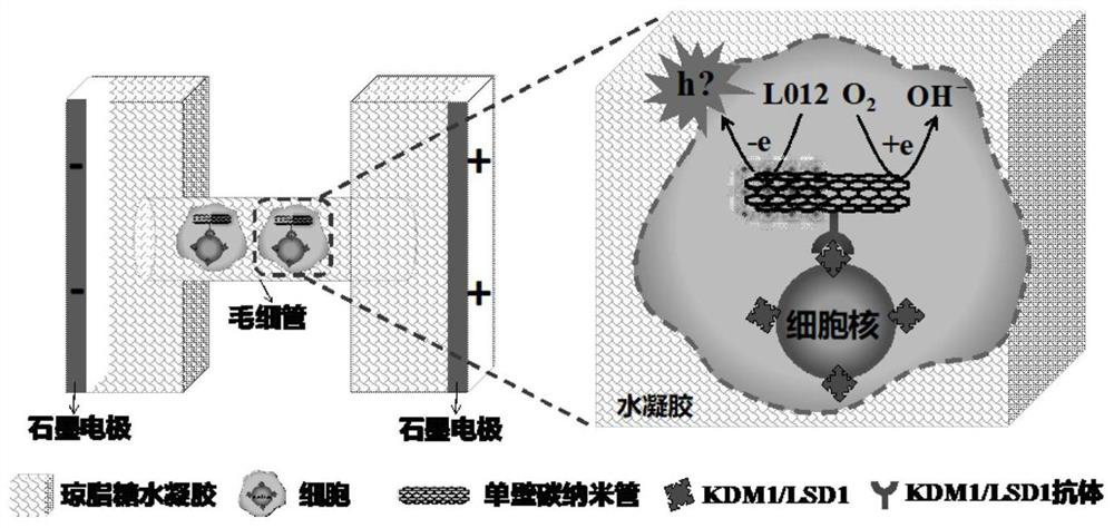
Radio chemistry visual analysis method for antigen in single cell
An analysis method and internal antigen technology, applied in the field of electrochemiluminescence, can solve the problems of poor prognosis, difficult operation, and hindering cell differentiation of acute myeloid leukemia
- Summary
- Abstract
- Description
- Claims
- Application Information
AI Technical Summary
Problems solved by technology
Method used
Image

Examples
Embodiment 1
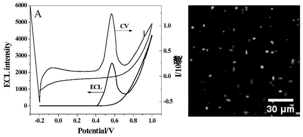
[0033] Example 1 Visualization experiment of single-armed carbon nanotubes in hydrogel
[0034] For initial visualization of individual SWCNTs using BPE-ECL, agarose containing SWCNTs-COOH (0.005% wt), L012 (luminol analog) (1 mM), and 10 mM phosphate buffered saline (PBS, pH 7.4) were hydrolyzed The glue is placed in a capillary (id. 0.6 mm). Through transmission electron microscopy, it is found that the length of a single SWCNTs-COOH is less than 500nm, and the width is less than 10nm. After the gel was formed in the capillary, the SEM image showed that the gel was a porous structure with a pore size of less than 70 μm, which was suitable for retaining a cell.
[0035] According to the results of this example, when only an electric field of 1kV / cm is applied, a resolvable ECL tube sheet can be observed in the capillary, see figure 2 b. As the electric field gradually increases from 1kV / cm to 1.2kV / cm, the ECL intensity of these points gradually increases. The dependence...
Embodiment 2
[0036] Example 2 Visual labeling of carcinoembryonic antigen on a single cell membrane
[0037] Nanotubes for Single-Cell BPE-ECL Imaging to Display Antigens on Cell Membranes. Carcinoembryonic antigen antibody (anti-CEA Ab) was labeled on SWCNTs to form anti-CEA Ab-SWCNTs complex, and then incubated with fixed MCF 7 cells (the cell membrane highly expresses CEA antigen), and then It was embedded in agarose hydrogel containing L012, and a voltage of 1000V was applied to the two feeding electrodes while recording ECL images. from image 3A-C It can be observed that the entire surface of a single cell produces a visible ECL spot, which overlaps well with the bright field cell. Furthermore, in order to rule out possible non-specific adsorption of SWCNTs, cells were incubated with SWCNTs without antibody, and ECL images were collected. The results showed that almost no ECL spots were observed on the cells, proving that the nonspecific adsorption of SWCNTs in cells was negligibl...
Embodiment 3
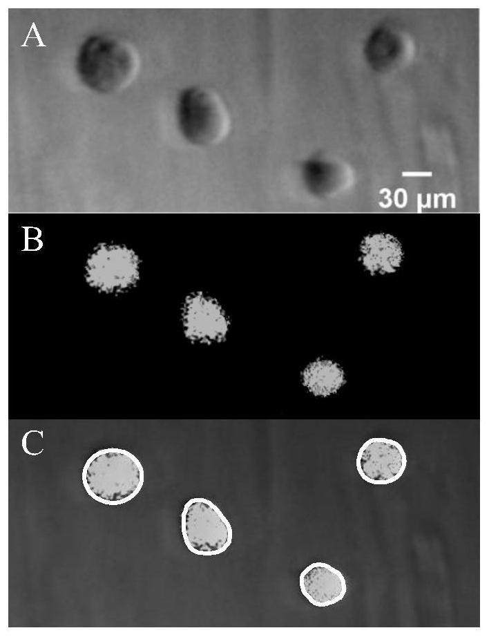
[0038] Example 3 Visual labeling of KDM1 / LSD1 protein on the nucleus
[0039] The present application further carried out an experiment of visually analyzing antigens in cells, namely: lysine-specific demethylase 1 (also known as KDM1 / LSD1). High expression of KDM1 / LSD1 can hinder cell differentiation and lead to poor prognosis in acute myeloid leukemia. First, cells were co-incubated with KDM1 / LSD1 antibody-modified SWCNTs, and then fixed with paraformaldehyde. Finally, the cells were permeabilized with Triton X-100 to facilitate the entry of L012 and buffer. Then, a voltage of 1 kV / cm was applied to the feed electrode, and an ECL image was recorded. like Figure 4 Overlay of brightfield and ECL images shown in A and B ( Figure 4 C) shows that ECL emission is concentrated near the nucleus in the cell, and is significantly different from the ECL visualization distribution of membrane proteins ( image 3 C). Fluorescent characterization of the position of the SWCNTs modi...
PUM
| Property | Measurement | Unit |
|---|---|---|
| length | aaaaa | aaaaa |
| pore size | aaaaa | aaaaa |
Abstract
Description
Claims
Application Information