Unlabeled proteomics detection method and device

A proteomics and detection method technology, applied in measurement devices, scientific instruments, biological testing, etc., can solve the problems of complex design, large sample size, and inability to continuously automatic sample injection, and achieve the effect of cost reduction and simple pretreatment.

Pending Publication Date: 2021-12-31
SHANGHAI JIAO TONG UNIV
View PDF9 Cites 2 Cited by
  • Summary
  • Abstract
  • Description
  • Claims
  • Application Information

AI Technical Summary

Problems solved by technology

The improved method based on non-labeled proteomics requires more samples, and the research cost based on TMT-labeled proteomics is more expensive
The proteomics of micro-samples using the microfluidic platform has obtained good identification results, the demand for cells is greatly reduced, and the proteomics detection of less than 100 cells can be realized, but the design is relatively complicated, and it is not easy to be used by other mass spectrometry experiments. At the same time, after the sample preparation of the microfluidic platform is completed, it is necessary to directly inject samples through the chromatographic column, which also puts forward high requirements for the operation of the liquid mass machine. Not only must there be a mature chromatographic column preparation system, but at the same time, each The sample time is relatively long, including the calibration, balance and sampling of the machine, and continuous automatic sampling is not possible

Method used


Image

  • Unlabeled proteomics detection method and device
  • Unlabeled proteomics detection method and device
  • Unlabeled proteomics detection method and device

Examples


Embodiment 1

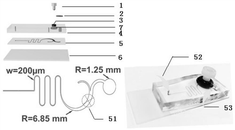
[0048] Example 1 Preparation of microfluidic system

[0049] (1) Pour PDMS and curing agent (Sylgard 184, Dow Corning) into a Petri dish on the surface of the silicon wafer at a ratio of 11:1, and bake at 85°C for 45 minutes.

[0050] (2) Use oxygen plasma to bond the nut to the PDMS solidified surface near the middle of the semicircular channel for 50 seconds.

[0051] (3) Pour PDMS and curing agent into a petri dish until the nuts are covered, and bake for 1 hour.

[0052] (4) Peel off the PDMS with nuts from the dish, and stick to the glass substrate after being treated with oxygen plasma for 50s.

[0053] (5) Punch holes at the inlet and outlet. The inlet and inner outlet adopt a punching machine with a diameter of 0.8mm, and the outer outlet adopts a punching machine with a diameter of 2.5mm.

[0054] (6) A filter membrane with a pore size of 20 μm is pasted on the top of the outlet.

[0055] (7) Treat the chip with 1% BSA for 1 h at room temperature to block the surf...

Embodiment 2293

[0056] Example 2 293T cell proteome sample preparation

[0057] (1) Prepare the target cells. After dilution, add the microfluidic system through the injection port and enter the sample pool.

[0058] (2) with 50mM NH 4 HCO 3 Chips were washed, replaced with PBS, and images of the cavity were taken through a Zeiss LSM 880, Germany microscope for accurate cell counts.

[0059] (3) The filter membrane is removed.

[0060] (4) The bolt is screwed into the nut to fix the cells in the exit mouth.

[0061] (5) Place the chip in an oven at 80°C for 30 minutes to denature cell proteins.

[0062] (6) Take 5 μL (0.1% DDM, 1mM TCEP, 2mM CAA dissolved in 50mM NH 4 HCO 3 ) to the outer export.

[0063] (7) The outer outlet was sealed with a sample bottle gasket and electrical tape, and incubated at 60° C. for 1 hour.

[0064] (8) Then add 5 μL of trypsin (total trypsin (w): protein (w) = 1:10) from the internal outlet, seal with the vial seal and electrical tape, and incubate overn...

Embodiment 3

[0067] Example 3 Separation, counting and proteome sample preparation of MCF7 cells

[0068] (1) The concentration of MCF7 cells was 5×10 4 cells / mL and 1×10 3 cells / mL and 5×10 respectively 4 The cells / mLWBCs are mixed, and 1mL / h of the microfluidic system is added through the injection port into the sample pool.

[0069] (2) Smaller cells and white blood cells flow out from the inner outlet, and MCF7 cells flow out from the outer outlet and pass through the filter membrane.

[0070] (3) with 50mM NH 4 HCO 3 Chips were washed, replaced with PBS, and images of the cavity were taken through a Zeiss LSM 880, Germany microscope for accurate cell counts.

[0071] (4) The filter membrane is removed.

[0072] (5) The bolts are screwed into the nuts to fix the cells in the exit mouth.

[0073] (6) Place the chip in an oven at 80°C for 30 minutes to denature cell proteins.

[0074] (7) Take 5 μL (0.1% DDM, 1 mM TCEP, 2 mM CAA dissolved in 50 mM NH4HCO3) and add it to the outer...


PUM

PropertyMeasurementUnit
diameteraaaaaaaaaa
diameteraaaaaaaaaa

Abstract

The invention discloses an unlabeled proteomics method for trace cell online separation tandem mass spectrometry detection, and relates to the field of single cell proteomics detection. The method realizes cell separation, sample preparation and tandem mass spectrometry detection by a microfluidic system. According to the invention, accurate counting of cells and pretreatment of trace biological samples are realized by establishing the microfluidic system, so that accurate quantification of protein information of a single cell is realized; and high-throughput proteomics detection of the CTCs is further carried out, and the drug resistance reaction of individuals to chemotherapy is monitored by utilizing proteomics information of the CTCs, so that the method has great significance in further guiding individualized medication.

Description

technical field [0001] The invention relates to a non-labeled proteomics method for online separation and tandem mass spectrometry detection of trace cells, in particular to a non-labeled proteomics detection method for realizing cell separation, sample preparation, and tandem mass spectrometry detection through a microfluidic system. Background technique [0002] In proteomics research, in addition to the sensitivity and high resolution of mass spectrometer hardware, sample pretreatment, mass spectrometry data acquisition and data analysis all have an impact on proteomics research. [0003] The sample pretreatment process is complex and needs to be optimized according to the specific experimental purpose to reduce the degradation and modification of the protein during the sample processing stage and release as many peptides as possible for the final mass spectrometry detection. Among them, micro-sample proteomics detection is a technical bottleneck. Samples such as flow cyt...

Claims


Application Information

Patent Timeline
Patent Type & Authority Applications(China)
IPC IPC(8): G01N30/72G01N30/06G01N33/68
CPCG01N30/72G01N30/06G01N33/6848
Inventor 丁显廷王丽萍朱大为
Owner SHANGHAI JIAO TONG UNIV
Why Eureka
Try Eureka