Skin wound healing preparation

A wound healing and skin technology, applied in the field of biology, to achieve the effect of reducing medical expenses, significant healing effect, and rapid healing effect

Pending Publication Date: 2022-02-22
DREAMTEC INTPROP LTD
View PDF0 Cites 0 Cited by
  • Summary
  • Abstract
  • Description
  • Claims
  • Application Information

AI Technical Summary

Problems solved by technology

Of the common treatments, decubitus deep bone and surgical treatment in

Method used


Image

  • Skin wound healing preparation
  • Skin wound healing preparation
  • Skin wound healing preparation

Examples


Embodiment 1

[0119] Example 1: Expression vector construction and host cell transformation

[0120] Construction and Design of Escherichia coli / Bacillus subtilis Expression Shuttle Vector

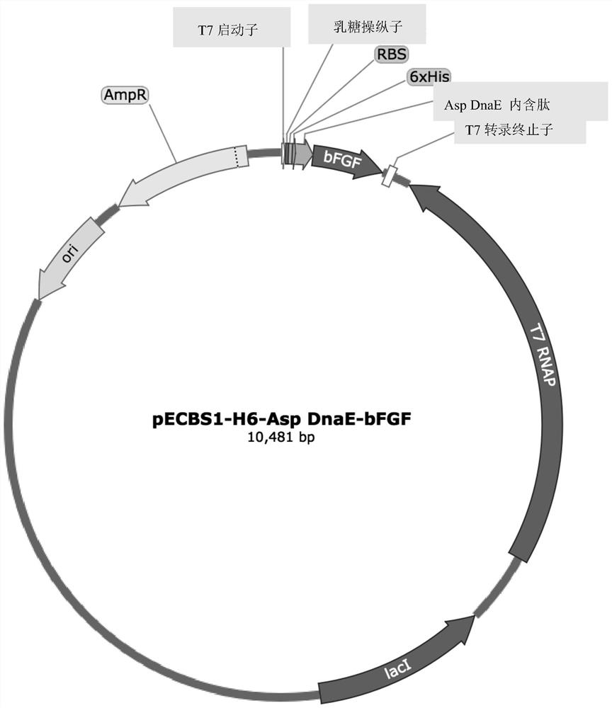
[0121] pRB374 and pBR322 were used as the starting vector of E. coli / Bacillus subtilis expression shuttle vector [14]. Specifically, pECBS1 was constructed by the following modification steps: first, pRB374 (5.9 kb) was digested with SalI and BglII; T7 ribonucleic acid polymerase-Lac promoter-LacI gene-LacI q The promoter-bleomycin resistance gene-partial neomycin resistance gene fragment (5.3kb) was substituted to form pECBSi vector. Then, the formed pECBSi vector and pBR322 vector were digested with EcoRI and BglI, respectively, and the pECBSi digested fragment was replaced with the fragment obtained by digesting pBR322 (4.3 kb), thereby forming the pECBS1 shuttle vector.

[0122] Construction of bFGF expression vector

[0123] The construction method of E. coli / Bacillus subtilis expression shut...

Embodiment 2

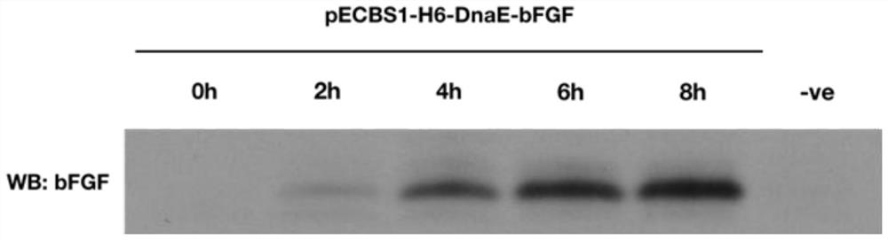
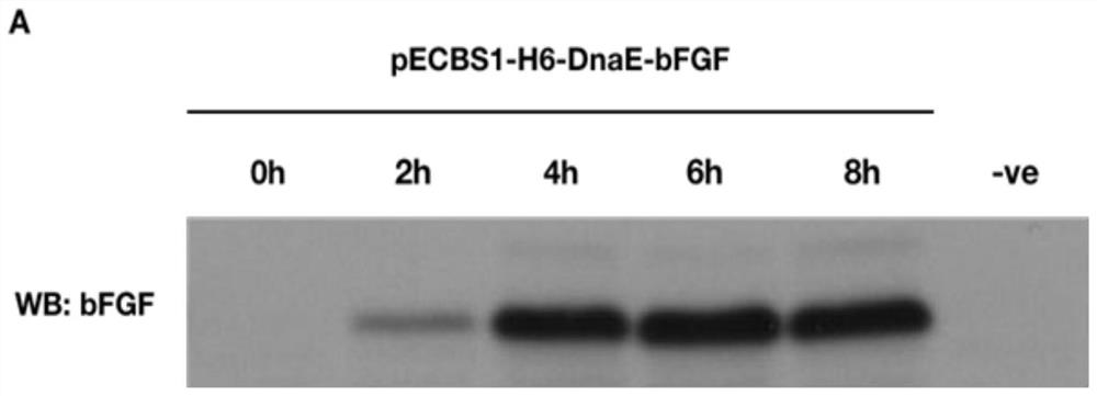
[0126] Example 2: Expression of bFGF

[0127] shake flask culture

[0128] B. subtilis transformants were grown at 37°C (250 rpm) in 200 ml 2x LB medium supplemented with 25 μg / ml kanamycin [15]. When the A600 value reached 1.0, IPTG was added at a final concentration of 0.2 mM, and then 1 ml of culture samples were collected at 3-hour intervals for bFGF expression analysis. The cell pellet was resuspended in 200 μl of resuspension buffer (50 mM Tris-Cl, 200 mM EDTA, pH 8.0) and incubated on ice for 5 minutes. The mixture was then treated with 120 μl of lysozyme solution (10 mg / mL) for 20 minutes at 37°C. Then 80 μl of lysis buffer (10 mM EDTA, 10% Triton X-100 and 50 mM Tris-Cl, pH 8.0) was added. The tube containing the solution was gently inverted and then centrifuged at 14,800 rpm for 5 minutes. Cell lysate samples were analyzed by Western blot for bFGF protein expression.

[0129] In order to successfully express the soluble bFGF protein, the inventors also tested ...

Embodiment 3

[0137] Example 3: Purification and structural determination of bFGF protein

[0138] Cation exchange chromatography and heparin-agarose chromatography were used to purify bFGF. First, the protein concentration of the eluted fractions was measured using a Nanodrop Microvolume spectrophotometer. In addition, eluted fractions with significant readings (approximately 1 mg / ml) were pooled and dialyzed against 0.1x PB. Afterwards, the purified bFGF band was obtained by electrophoresis on a 10% SDS-PAGE gel stained with Coomassie brilliant blue R-250. The band containing bFGF protein in the SDS-PAGE gel was recovered for subsequent analysis by LC-MS.

[0139] The results of Western blot analysis showed that the soluble bFGF protein extracted from the lysate had the same molecular weight as the bFGF protein purchased from Thermofisher Scientific ( FIG. 4 ). Purified bFGF protein samples were subjected to N-terminal and C-terminal protein sequencing and MALDI-TOF mass determination ...


PUM


Abstract

The invention discloses a skin wound healing preparation, and the effective components of the skin wound healing preparation comprise an epidermal growth factor and/or a fibroblast growth factor, the epidermal growth factor and/or the fibroblast growth factor are/is obtained by culturing transformed host cells; the transformed host cell comprises an expression vector, the expression vector comprises a nucleic acid construct, and the nucleic acid construct comprises an insert, the insert comprises, from the 5'end to the 3'end, a polynucleotide sequence encoding an oligopeptide affinity tag, a trans-spliced intein derived from Anabaena sp., and an exogenous polypeptide, wherein the oligopeptide affinity tag is used as the N-terminal exon peptide of the trans-spliced intein, and the exogenous polypeptide is used as the C-terminal exon peptide of the trans-spliced intein. Through local application of the skin wound healing preparation, the cure rate of diabetic foot ulcers, bedsores and hard-to-heal wounds is greatly increased.

Description

technical field [0001] The invention relates to the field of biology, in particular to a skin wound healing preparation. Background technique [0002] Diabetic foot ulcers and bedsores are difficult-to-heal wounds that can deteriorate and become life-threatening if left untreated. The general treatment for diabetic foot ulcers is antibiotic administration, necrotic debridement, and limb amputation. The efficacy of antibiotics and debridement is low, and amputation is an irreversible procedure with potentially high risk of death. The cost of amputation is also expensive, ranging from HK$200,000 to HK$600,000. [0003] Bed sores are a common ailment of the elderly, where prolonged pressure on local body tissues can block blood circulation and make wound healing difficult. Among the common treatment methods, decubitus deep bone and surgical treatment (including debridement of necrotic tissue) proved ineffective. Contents of the invention [0004] In order to solve the pro...

Claims


Application Information

Patent Timeline
IPC IPC(8): A61K38/18A61P17/02
CPCA61K38/1808A61K38/1825A61P17/02A61K2300/00
Inventor 钟树根邝纬阳林庭匡
Owner DREAMTEC INTPROP LTD
Why Eureka
Try Eureka