Preparation and application of epilepsy animal model
A technology of epilepsy animals and animal models, applied in the direction of DNA / RNA fragments, recombinant DNA technology, hydrolytic enzymes, etc., can solve the problems of reduced efficiency, lack of refractory epilepsy in patients with simulated CDKL5 syndrome, and poor efficacy of antiepileptic drugs
- Summary
- Abstract
- Description
- Claims
- Application Information
AI Technical Summary
Problems solved by technology
Method used
Image
Examples
Embodiment 1
[0103] Example 1 Preparation of epileptic seizure mice
[0104] Age-appropriate female Cdkl5 flox / flox The mice were mated with male mice specifically expressing Cre recombinase, and the toes of the pups were numbered seven days after birth, and the tails of about 3 mm were taken for genotype identification. Submerge the mouse tail in 150ul of mouse tail lysis solution (containing 2ul 5% proteinase K solution), incubate overnight at 55°C; place the centrifuge tube containing the lysis solution on a metal bath at 95°C and heat for 15min; cool to After room temperature, centrifuge at 12000rpm / min for 5min; take the supernatant for PCR identification of genotype. Prepare the PCR reaction system, the formula is as follows:
[0105]
[0106] The PCR cycling conditions were:
[0107] 95°C for 15s, 60°C for 15s, 72°C for 45s, for 35 cycles.
[0108] The PCR product and 2x sample buffer were mixed and added to 1.5% agarose gel, electrophoresed in a constant voltage electric fie...
Embodiment 2
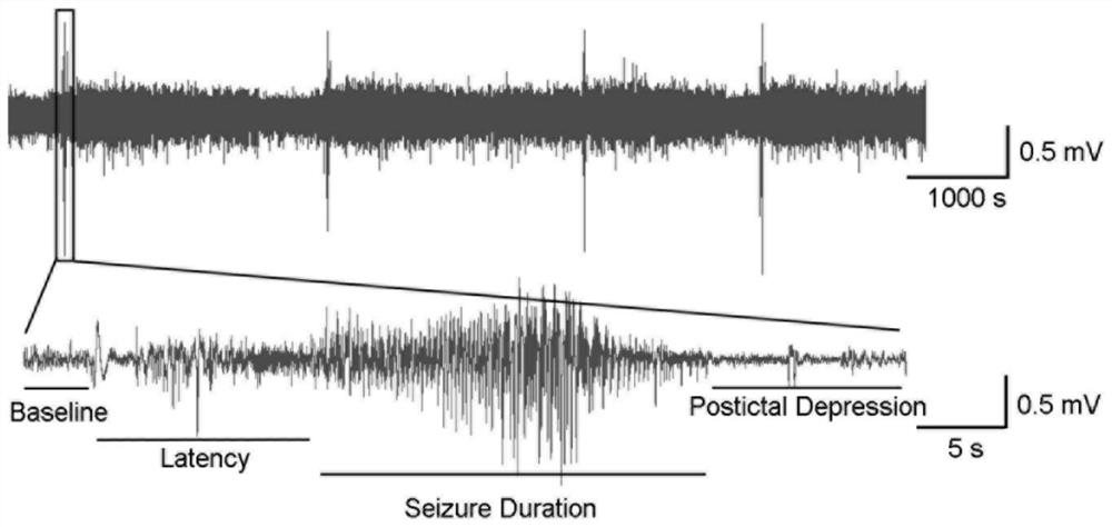
[0112] Example 2 Accurately determine the frequency of epileptic seizures in transgenic mice by means of EEG recording
[0113] The self-made electrode was installed on the mouse pia mater by surgery. During the operation, the mouse was anesthetized with 1.5% isoflurane and placed above the stereotaxic position so that the skull was in a horizontal position. The self-made electrode consists of two stainless steel screws (diameter 1mm), which are used as EEG recording electrodes, and the position of insertion into the skull is determined according to the mouse brain atlas (the coordinates of the bregma as the origin: front-to-back +1.0mm, mid-lateral +1.5mm and front-to-back- 3.2mm, mediolateral +1.5mm). Two insulated silver wires (Coonerwire, #AS633) were used as EMG recording electrodes, which were placed in the trapezius muscles on both sides. The electrodes are attached to miniature connectors and secured to the skull with dental cement. The mice's skin was then closed wi...
Embodiment 3
[0116] The method of embodiment 3 Timm staining determines that CDKL5-related epilepsy has occurred in mice
[0117] Mossy fiber sprouting in the dentate gyrus of the hippocampus is a characteristic morphological alteration in limbic epilepsy patients and mice Timm staining was performed on the hippocampal slices of CDKL5 conditional knockout mice with epilepsy, and it was found that the axon fibers of granule cells were distributed in bands in the molecular layer of the dentate gyrus ( Figure 5 , B-B'), while the control mice without epilepsy ( Figure 5 , A-A', C-C', E-E') and VGAT-Cre-mediated cKO mice ( Figure 5 , D-D'), CDKL5 whole body knockout mice ( Figure 5 , F-F') There was no phenomenon of granule cell axon fibers projecting to the inner molecular layer of the dentate gyrus. The statistical results also showed that the ectopic projection of granulosa cells caused by the conditional knockout of CDKL5 mediated by Emx1-Cre had a statistically significant differen...
PUM
 Abstract
Description
Claims
Application Information
 - R&D
- Intellectual Property
- Life Sciences
- Materials
- Tech Scout
- Unparalleled Data Quality
- Higher Quality Content
- 60% Fewer Hallucinations
Browse by: Latest US Patents, China's latest patents, Technical Efficacy Thesaurus, Application Domain, Technology Topic, Popular Technical Reports.
© 2025 PatSnap. All rights reserved.Legal|Privacy policy|Modern Slavery Act Transparency Statement|Sitemap|About US| Contact US: help@patsnap.com


