Bionic body surface tissue and step-by-step construction method thereof

A construction method and body surface technology, applied in prosthetics, medical science, bone implants, etc., can solve the problems of insufficient matrix and microstructure, insufficient mechanical properties, etc., to meet the needs of clinical applications, improve mechanical strength, get handy effects

Active Publication Date: 2022-04-29
PLASTIC SURGERY HOSPITAL CHINESE ACAD OF MEDICAL SCI
View PDF3 Cites 0 Cited by
  • Summary
  • Abstract
  • Description
  • Claims
  • Application Information

AI Technical Summary

Problems solved by technology

Therefore, the core problem of insufficient mechanical propert

Method used


Image

  • Bionic body surface tissue and step-by-step construction method thereof
  • Bionic body surface tissue and step-by-step construction method thereof
  • Bionic body surface tissue and step-by-step construction method thereof

Examples


Embodiment 1

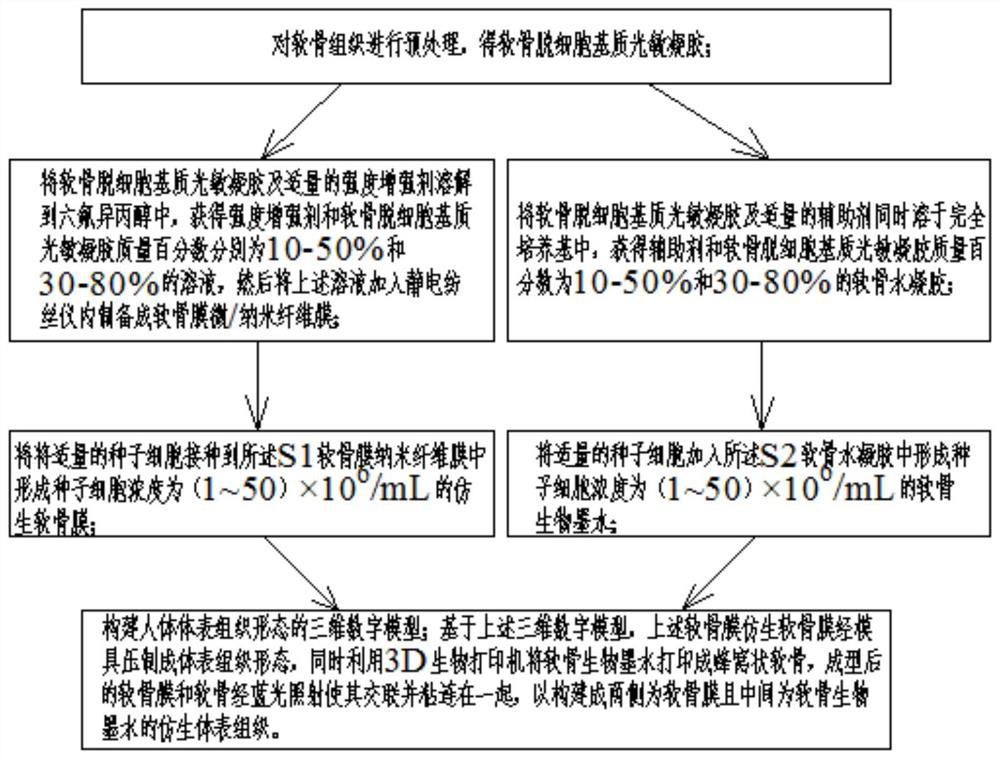
[0053] Such as Figure 1 to Figure 13 As shown, this embodiment provides a step-by-step construction method of bionic body surface tissue, including the following specific steps:

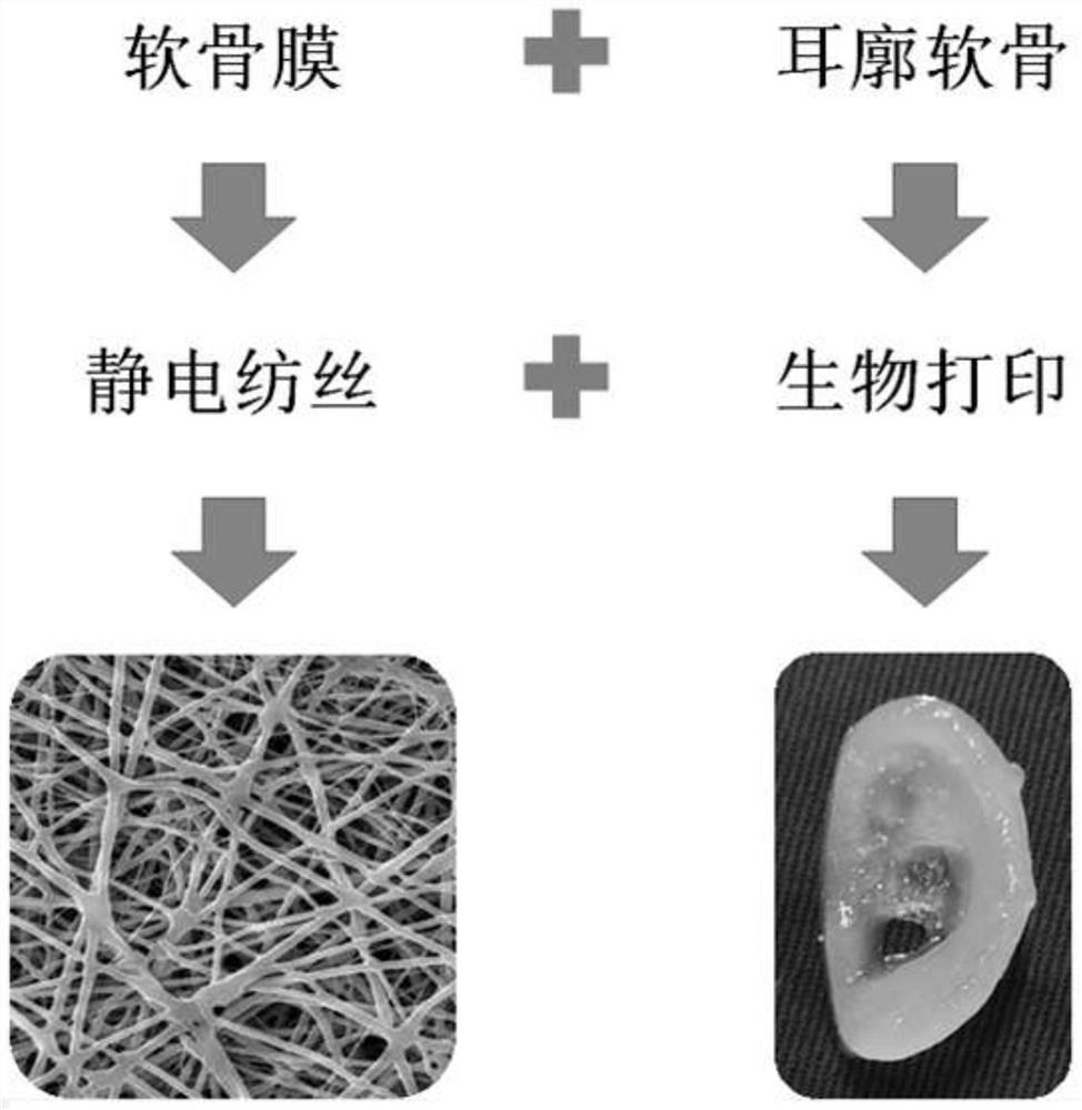
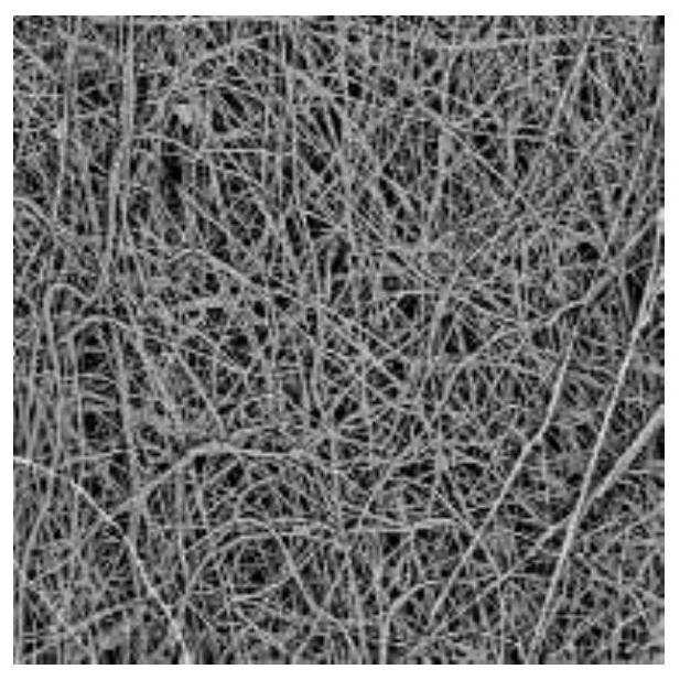
[0054] S1: The cartilage decellularized matrix photosensitive gel and an appropriate amount of strength enhancer are dissolved in hexafluoroisopropanol, and the mass percentages of the strength enhancer and cartilage decellularized matrix photosensitive gel are 10-50% and 30-80% respectively solution, and then add the above solution into the electrospinning apparatus to prepare a perichondrium micro / nano fiber membrane;

[0055] S2: Dissolving cartilage decellularized matrix photosensitive gel and appropriate amount of auxiliary agent in complete medium at the same time to obtain cartilage hydration with the mass percentage of auxiliary agent and cartilage acellular matrix photosensitive gel being 10-50% and 30-80% glue;

[0056] Among them, one or more components of basic fibroblast growth factor...

Embodiment 2

[0061] On the basis of Example 1, in this example, before S1, S0 is also included: pretreating the cartilage tissue to obtain a cartilage decellularized matrix photosensitive gel. Simple preparation process, providing biomimetic raw material cartilage acellular matrix photosensitive gel for perichondrium and cartilage bioink

Embodiment 3

[0063] On the basis of Example 2, in this example, the cartilage tissue in the S0 is one or more of ear cartilage, articular cartilage, costal cartilage, scapular cartilage, and meniscus. The method is convenient to obtain, and is beneficial to the preparation of bionic reconstructed body surface tissue.

[0064] It should be noted that the above-mentioned cartilage tissue is usually pig cartilage tissue, which can be purchased in the market and can be industrialized.


PUM

PropertyMeasurementUnit
Particle sizeaaaaaaaaaa

Abstract

The invention relates to a bionic body surface tissue and a step-by-step construction method thereof, and the method comprises the following specific steps: preparing cartilage acellular matrix photosensitive gel and an auxiliary agent into a micro/nano fiber membrane by using an electrostatic spinning instrument, and preparing a bionic cartilage membrane; dissolving the cartilage acellular matrix photosensitive gel and an auxiliary agent in a complete culture medium to form cartilage hydrogel; adding the seed cells into the cartilage hydrogel to form biological ink; and constructing a bionic body surface tissue by using the bionic cartilage membrane and the biological printed cartilage. The structure and composition characteristics of the cartilage membrane-containing body surface tissue are simulated, an acellular matrix material is used, the electrostatic spinning and biological printing technologies are combined to realize dual biomimetic of the composition and the structure, different functional cells are used to realize matrix biomimetic, and the body surface cartilage tissue with the cartilage membranes on the two sides and the cartilage tissue in the middle is constructed step by step. And mechanical bionics are achieved through structure and component bionics, the mechanical strength of constructors is improved, and the clinical application requirements of body surface tissue reconstruction are met.

Description

technical field [0001] The invention relates to the technical field of three-dimensional bioprinting and tissue and organ repair and reconstruction, in particular to a bionic body surface tissue and a step-by-step construction method thereof. Background technique [0002] Ear cartilage defects caused by congenital diseases, trauma, etc. seriously affect the physical and mental health of patients, and the problem of repair and reconstruction has brought a heavy burden to families and society. Cartilage tissue engineering provides a new strategy for auricle reconstruction. The first international clinical breakthrough of tissue engineering auricle has been successfully achieved in the early stage by using polyglycolic acid / polylactic acid (PGA / PLA) and chondrocytes. However, long-term follow-up found that the three-dimensional structure of the reconstructed auricle gradually blurred, atrophied, deformed, and collapsed, and the long-term clinical effect did not meet expectation...

Claims


Application Information

Patent Timeline
IPC IPC(8): A61L27/18A61L27/22A61L27/36A61L27/38A61L27/50A61L27/56A61F2/28A61F2/18
CPCA61L27/3633A61L27/3687A61L27/3695A61L27/3817A61L27/3834A61L27/222A61L27/227A61L27/18A61L27/56A61L27/50A61F2/28A61F2/18A61F2/30756A61F2/30942A61F2002/183A61F2002/30762A61F2002/30943A61F2002/30985A61L2430/14A61L2430/22A61L2430/24C08L67/04C08L89/00
Inventor 刘霞贾立涛刘文帅蒋海越
Owner PLASTIC SURGERY HOSPITAL CHINESE ACAD OF MEDICAL SCI
Why Eureka
Try Eureka