Preparing process for optical fibre bio-sensor of acetylcholinesterase

A technology of acetylcholinesterase and biosensor, which is applied in the field of preparation of acetylcholinesterase optical fiber biosensor, can solve the problems of non-reusable use, long time, complicated process, etc., and achieve the effect of mechanization, accurate identification and simple operation

Inactive Publication Date: 2004-08-11
NANJING MEDICAL UNIV
View PDF0 Cites 1 Cited by
  • Summary
  • Abstract
  • Description
  • Claims
  • Application Information

AI Technical Summary

Problems solved by technology

However, this method needs to keep the patient awake, and is limited to fresh injuries, individual differences are large, and it is impossible to distinguish whether the muscle contraction caused by stimulation is the result of nerve conduction or current diffusion.
Therefore, the clinical application value is not large
[0005] 3. Radiobiochemical method: use radioactive isotope to label the substrate of the enzyme, and then measure the activity of the enzyme according to the radioactive product produced by the enzyme reaction, and infer the nature of the nerve bundle, but this method involves isotope technology, high equipment requirements, and radiation The substance is harmful to both the patient and the operator, so it is not very practical
However, there are problems with this method: (1) the staining time is long (about 50 minutes), and the process is complicated; (2) two nerve fragments need to be excised, and in situ staining cannot be performed
However, the problem with this method is that the sensor needle must be replaced after each nerve bundle is detected, and cannot be reused. It still takes a few minutes to detect a nerve bundle, which is a bit too long

Method used


Image

  • Preparing process for optical fibre bio-sensor of acetylcholinesterase
  • Preparing process for optical fibre bio-sensor of acetylcholinesterase
  • Preparing process for optical fibre bio-sensor of acetylcholinesterase

Examples


Embodiment 1

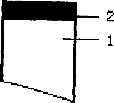
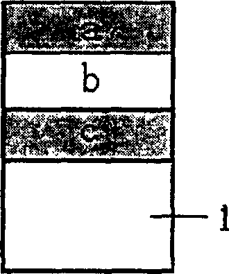
[0031] Embodiment 1: The diameter of the Y-shaped optical fiber 1 is 1 mm, and the sensing film 2 has a three-layer structure. The first layer is a Nafion polymer membrane a, the middle is a choline oxidase layer b, and the third layer is a polymer layer modified with an oxidation indicator c, and the oxygen indicator is Tris(2,2'-bipyridyl)ruthenium(II )chlorideHexahydrate. The sensing film 2 is fixed to the surface of the optical fiber head of the Y-shaped optical fiber 1 by means of covalent bonding.

Embodiment 2

[0032] Embodiment 2: The diameter of the Y-shaped optical fiber 1 is still 1 mm, and the sensing film 2 has a two-layer structure. Choline oxidase membrane and oxygen indicator membrane. The oxygen indicator membrane is 5mm(bpy) 3 2+ 1.25wt% Nafion ethanol solution was dropped onto the end face of the optical fiber and dried at room temperature. Choline oxidase solution is Nafion solution with 90%

[0033]Ethanol was diluted to a final concentration of 1%, and 3mg of choline oxidase (10 units / mg) was added to 50ul% Nafion solution. The prepared choline oxidase liquid is dropped onto the surface of the optical fiber to dry to prepare a sensing film.

Embodiment 3

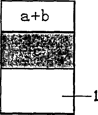
[0034] Embodiment 3: The Y-shaped optical fiber 1 has a diameter of 1 mm, and the sensing film 2 has a single-layer structure. The solutions of oxygen indicator C, choline oxidase b and PDMS polymer material a are mixed and condensed to form a monolayer film. The above-mentioned single-layer sensing film 2 is fixed to the surface of the optical fiber head of the Y-shaped optical fiber 1 by means of adsorption.

[0035] According to the acetylcholinesterase optical fiber sensor that embodiment 1 to embodiment 3 make, combine light source 3, detector 4, computer 5 and image display 6 (such as Figure 5 ), the properties of the nerve bundles of the nerve trunk 7 can be quickly, intuitively and accurately determined. The light source can be laser, light-emitting diode, mercury lamp, etc. When using a light source with a wide spectrum such as a mercury lamp, use the corresponding color filter to obtain the excitation light of the required wavelength, and the wavelength selection ...


PUM

PropertyMeasurementUnit
diameteraaaaaaaaaa

Abstract

An optical fibre bio-sensor for distriminating the nature of peripheral nerve fibres is composed of optical fibre and sensing membrane which is fixed to the end of optical fibre by co-valent binding or physical adsorption. Said sensing membrane is prepared from high-molecular film containing oxygen indicator, choline oxidase and high-molecular membrane by one of three methods. Its advantages include no need of cutting nerve fibre, high speed and correctness and no poison and no wound to human body.

Description

1. Technical field [0001] The invention relates to a method for preparing a sensor for detecting the properties of peripheral nerve bundles, in particular to a method for preparing an acetylcholinesterase optical fiber biosensor. 2. Background technology [0002] In the prior art, there are mainly the following methods for judging the properties of nerve bundles: [0003] 1. Anatomical method: The localization map of the nerve bundles in the nerve trunk has been studied. Many scholars have analyzed the peripheral nerves (such as the median nerve, radial nerve, ulnar nerve trunk, sciatic nerve trunk, etc.) respectively, and observed the nerve trunk. The position and shape of the number of bundles were measured, and the local location map of the nerve bundles was drawn on the nerve section. However, in clinical practice, the two ends are prone to distortion after nerve injury, which changes the original normal anatomical position. Therefore, clinical application restricted. ...

Claims


Application Information

Patent Timeline
Patent Type & Authority Patents(China)
IPC IPC(8): C12Q1/44
Inventor 曹晓建陆祖宏王宏柴雪挺贾可
Owner NANJING MEDICAL UNIV
Why Eureka
Try Eureka