
Preparing method of nerve tissue engineering tube type bracket and special mould for the same

A nerve tissue, tubular technology, applied in the field of biomedical materials and tissue engineering, can solve the problems of unable to maintain the structure, difficult to preset cells, unsuitable for small-bore catheters, etc., to achieve the effect of promoting nerve regeneration

Inactive Publication Date: 2007-03-14
TSINGHUA UNIV
View PDF10 Cites 2 Cited by
  • Summary
  • Abstract
  • Description
  • Claims
  • Application Information

AI Technical Summary

Problems solved by technology

Chinese patent application 00810000.4 provides a nerve conduit made of polyester material. The lumen is filled with collagen hydrochloric acid solution and freeze-dried to obtain a collagen content with fine fibrils. However, this method is difficult to obtain a multi-channel structure with axial penetration ; Chinese patent application 02113103.1 puts chitosan fibers into chitosan tubes to prepare pure chitosan nerve guides; Chinese patent application 02105864.4 places folded and curled porosity with concave-convex surfaces in hollow tubes made of polyester materials Polyester films are used to increase the surface area of ​​the cavity to make multi-channel nerve regeneration catheters. However, the contents of the above-mentioned catheters are in a free state, which cannot maintain a stable structure, and it is difficult to preset cells.
However, the catheter-guided axonal regeneration ability of random structures made by particle leaching pore-making technology is unreliable, and hot-extrusion technology cannot add therapeutic drugs or factors to the matrix material, because the mechanical processing and heat treatment process will make them denature or degrade, and current hot extrusion techniques are not suitable for making small-bore catheters suitable for nerve repair
Rapid prototyping technology simulates the principle of inkjet printers to print three-dimensional organs, but due to the limitation of materials, some biological materials (such as collagen and chitosan) cannot be applied to this method, and drug factors can not tolerate such a processing process. limit its application

Method used


Image

  • Preparing method of nerve tissue engineering tube type bracket and special mould for the same
  • Preparing method of nerve tissue engineering tube type bracket and special mould for the same
  • Preparing method of nerve tissue engineering tube type bracket and special mould for the same

Examples


preparation example Construction

[0022] The preparation method provided by the present invention is as follows:

[0023] (1) Dip the PTFE-coated stainless steel rod core mold (1~5mm in diameter) into 2~4% (w / v) chitosan solution (prepared with 0.2mol / L acetic acid solution), pull it out, Freeze, then immerse, pull out, and freeze at least 3 times, then freeze at -20°C to -96°C for 12 to 48 hours, and then place it in a freeze dryer for freeze drying. Soak the dried chitosan round tube with a core mold in a 2% (w / v) sodium hydroxide solution for 10-30 minutes, take it out, rinse with deionized water to neutrality, and then immerse it in pH=7.2-7.4 in phosphate buffer for 10-30 minutes. Use filter paper to absorb moisture on the surface of the chitosan tube and dry it naturally. Take out the stainless steel rod core mold, and finally obtain a semi-permeable chitosan round tube with a wall thickness of 0.4-1.0 mm.

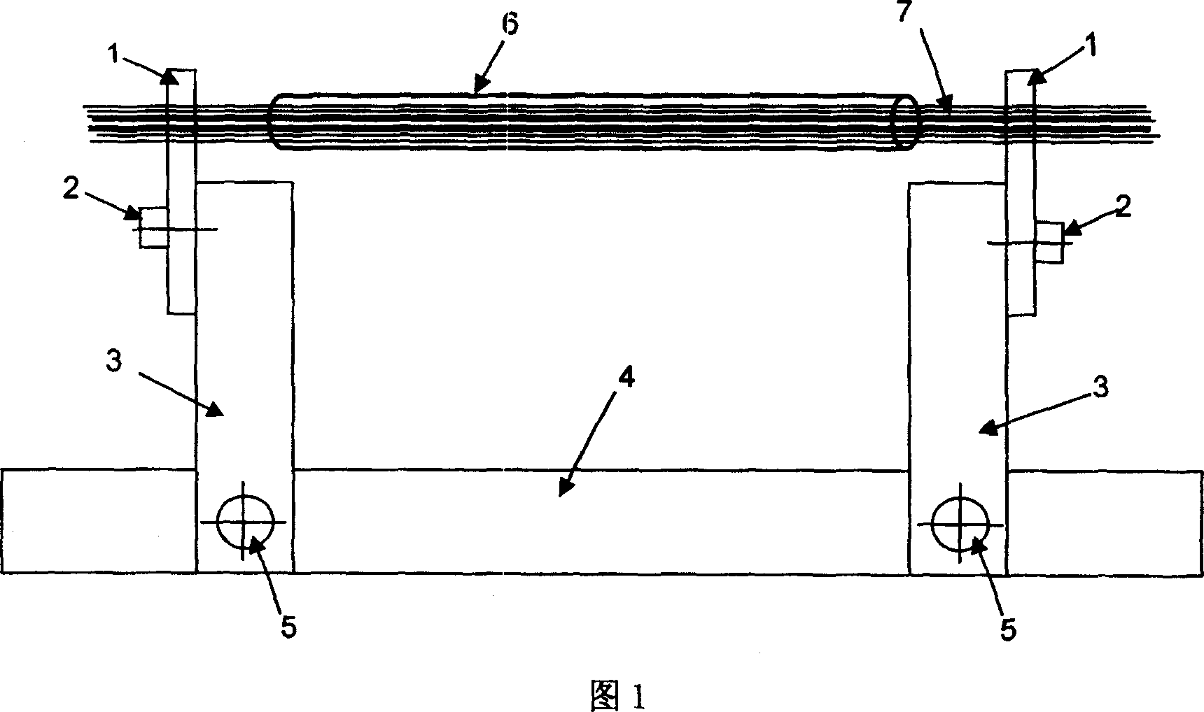
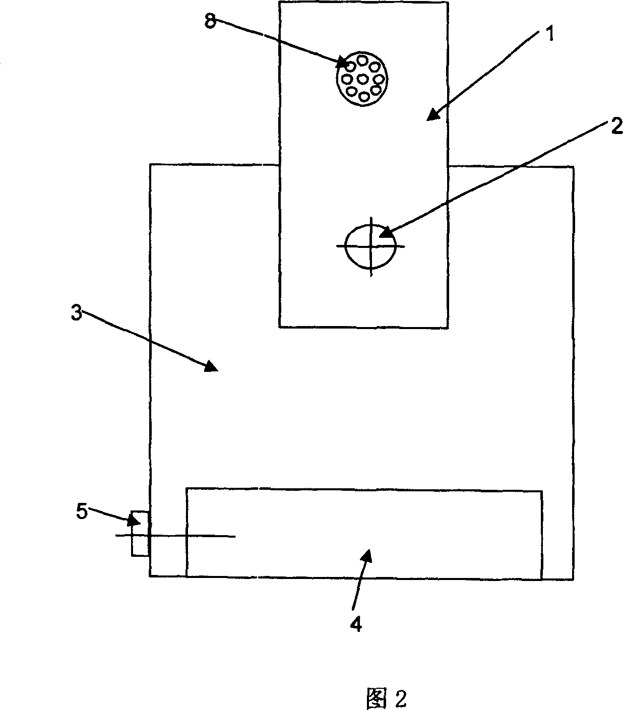
[0024] (2) The mold shown in Figure 1 can be used to prepare the multi-channel matrix. The mold inclu...

Embodiment 1

[0027] (1) Dip the stainless steel rod core mold with a diameter of 1mm PTFE-coated into 2% (w / v) chitosan solution (prepared with 0.2mol / L acetic acid solution), pull out, freeze, then immerse and pull out Take out and freeze, repeat 3 times, then freeze at -20°C for 12 hours, then place it in a freeze dryer for freeze drying. Soak the dried chitosan round tube with a core mold in a 2% (w / v) sodium hydroxide solution for 10 minutes, take it out, rinse with deionized water to neutral, and then soak in pH= 7.2 ~ 7.4 phosphate buffer for 10 minutes. Use filter paper to absorb the moisture on the surface of the chitosan tube and dry it naturally. Take out the stainless steel rod core mold, and finally obtain a semi-permeable chitosan round tube with a wall thickness of 0.4 mm.

[0028] (2) As shown in the figure, the chitosan tube 6 with a length of 1cm is placed between the two steel needle fixing plates 1 on the special mold (Figure 1), and 7 stainless steel needles with a diameter...

Embodiment 2

[0030] (1) Dip the stainless steel rod core mold with a diameter of 5mm PTFE coated into 4% (w / v) chitosan solution (prepared with 0.2mol / L acetic acid solution), pull out, freeze, then immerse and pull out Take out and freeze, repeat 5 times, then freeze at -196°C for 48 hours, then place it in a freeze dryer for freeze drying. Soak the dried chitosan tube with a core mold in a 2% (w / v) sodium hydroxide solution for 30 minutes, take it out, rinse with deionized water to neutral, and then immerse it in pH= 7.2 ~ 7.4 phosphate buffer for 30 minutes. Use filter paper to absorb the moisture on the surface of the chitosan tube and dry it naturally. Take out the stainless steel rod core mold, and finally obtain a semi-permeable chitosan round tube with a wall thickness of 1.0 mm.

[0031] (2) As shown in the figure, the chitosan tube 6 with a length of 10cm is placed between the two steel needle fixing plates 1 on the special mold (Figure 1), and 50 stainless steel needles 7 with a dia...


PUM

PropertyMeasurementUnit
thicknessaaaaaaaaaa
pore sizeaaaaaaaaaa

Abstract

Disclosed is a nervous tissue engineering tubular supporting stand and method for making same, wherein the nervous tissue engineering tubular supporting stand comprises a chitosan pipe wall and biological source filling base material with axial multiple passages, and the method consists of preparing semipermeable chitosan hollow pipe with 1-5mm of inside diameter, pouring large biological molecule solvent such as chitosan, collagen or gelatine, and utilizing special-purpose die arrangement and lyophilization technology. The obtained multiple-pass nerve channel with bionic construction is beneficial for cell adhesion, migration and leading of neuraxon directional growth, and suitable of the renovation and regeneration of nerve damages.

Description

Technical field [0001] The invention relates to a nerve tissue engineering tubular scaffold and a preparation method thereof, in particular to a nerve catheter with an axial multi-channel specific microstructure matrix, a preparation method and a special mold thereof, and belongs to the technical field of biomedical materials and tissue engineering. Background technique [0002] Peripheral nerve injury is very common in clinic. Once the nerve is damaged, the sensory and motor function of the corresponding part will be affected or even lost. The current clinically effective treatment method is autologous nerve transplantation. However, the source of autologous nerves is limited, and the function of the donor site is impaired. People try to use skeletal muscle and vein instead of autologous nerve transplantation, with little success. With the development of medicine, biology and tissue engineering, people consider and try to replace autologous nerve grafts with artificial nerve cat...

Claims


Application Information

Patent Timeline
Patent Type & Authority Patents(China)
IPC IPC(8): A61F2/04A61L27/00A61L27/20A61L27/24
Inventor 敖强王爱军张秀芳公衍道赵南明
Owner TSINGHUA UNIV
Learn More