Immune chromatography with fluorescent rare earth nanometer particle as marker and detecting testing paper strip
A nanoparticle and immunochromatographic technology, applied in biological testing, fluorescence/phosphorescence, measurement devices, etc., can solve the problems of difficult recording and storage of test results, low detection sensitivity, and difficult quantitative detection, and achieve easy operation and high sensitivity. , the effect of high sensitivity
- Summary
- Abstract
- Description
- Claims
- Application Information
AI Technical Summary
Problems solved by technology
Method used
Image
Examples
Embodiment 1
[0044] Example 1: Preparation of HBsAg Rapid Detection Test Strips
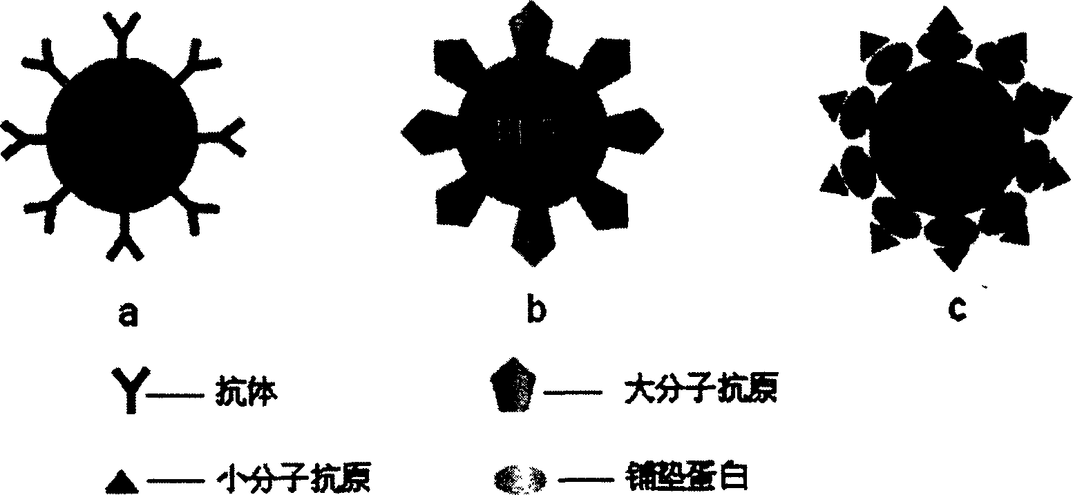
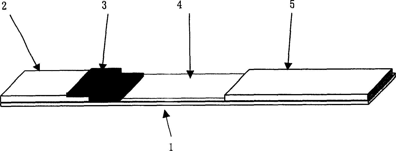
[0045] In this embodiment, HBsAg is used as the detection object, and the bonded fluorescent rare earth complex nanoparticles with a diameter of about 50nm and nitrocellulose chromatography membrane with amino groups on the surface are taken as examples. Through labeling, coating and assembly of various components, The finished product of the immunochromatographic detection test strip using fluorescent rare earth complex nanoparticles as markers is prepared.
[0046] (1) Labeling of anti-HBsAg monoclonal antibody
[0047] Method 1: Directional Markup
[0048] Take 500 μL (3.6 mg / mL) of affinity-pure anti-HBsAg monoclonal antibody, dialyze against 0.01 M sodium acetate buffer (pH5.2) at 4 °C for 6 h; then add NaIO 4 , to oxidize the antibody. After 20 minutes, dialyze against 0.01M pH5.2 sodium acetate buffer again; then add 500 μL (18 mg / mL) of BHHCT-Eu particles with amino groups on the surface, mix well,...
Embodiment 2
[0062] Example 2: Detection of biotin by biotin-nucleophile system simulation competition method
[0063] This example combines streptavidin mimetic capture antibody with biotinylated Tb 3+ The complex nanoparticles were used as markers, and the biotin was simulated by the competition method. The particle diameter used in the example is about 50nm.
[0064] (1)Tb 3+ Biotinylation of Complex Nanoparticles
[0065] method one:
[0066]Take 500 μL of BPTA-Tb particles with amino groups on the surface to be labeled (suspended in absolute ethanol), add a little biotin-mPEG-SPA, dissolve, mix well, and react at room temperature for 3 h; then, add absolute ethanol and 50 mM tris 7.8 The particles were washed 3 times with buffer, and then washed with 500 μL 50 mM tris 7.8 (containing 0.9% NaCl, 0.2% BSA, 0.1% NaN 3 ) for suspension.
[0067] Method Two:
[0068] Take 500 μL of BPTA-Tb particles with amino groups on the surface to be labeled (suspended in ultrapure water), treat...
Embodiment 3
[0077] Embodiment 3: One-step multi-inspection detection
[0078] The one-step multi-detection project was simulated with the HBsAg detection system and the biotin-nucleophile system. The HBsAg detection system of the first detection zone uses a pair of monoclonal antibodies at different sites as the capture antibody and the labeled antibody, in which the labeled antibody and Eu 3+ The complex nanoparticles are cross-linked and detected by the sandwich method. The biotin-nucleophile system of the second detection zone uses nucleophilic analog capture antibody to biotinylated Tb 3+ The complex nanoparticles were used as markers, and the biotin was simulated by the competition method. Rabbit anti-mouse IgG was used as the detection band to capture the antibody. The marker pad also contains anti-HBsAg monoclonal antibody labeled Eu 3+ Complex nanoparticles and biotin-labeled Tb 3+ complex nanoparticles.
[0079] (1) Labeling of anti-HBsAg monoclonal antibody
[0080] Same ...
PUM
| Property | Measurement | Unit |
|---|---|---|
| diameter | aaaaa | aaaaa |
| Sensitivity | aaaaa | aaaaa |
| Sensitivity | aaaaa | aaaaa |
Abstract
Description
Claims
Application Information
 - R&D
- Intellectual Property
- Life Sciences
- Materials
- Tech Scout
- Unparalleled Data Quality
- Higher Quality Content
- 60% Fewer Hallucinations
Browse by: Latest US Patents, China's latest patents, Technical Efficacy Thesaurus, Application Domain, Technology Topic, Popular Technical Reports.
© 2025 PatSnap. All rights reserved.Legal|Privacy policy|Modern Slavery Act Transparency Statement|Sitemap|About US| Contact US: help@patsnap.com


