Use of human phylaxin-1,2,3 protein in cancer detecting molecular mark
A technology of human defensin and detection label, applied in biological testing, material inspection products, etc., to reduce the cost of detection and diagnosis and improve the accuracy
- Summary
- Abstract
- Description
- Claims
- Application Information
AI Technical Summary
Problems solved by technology
Method used
Image
Examples
Embodiment 1
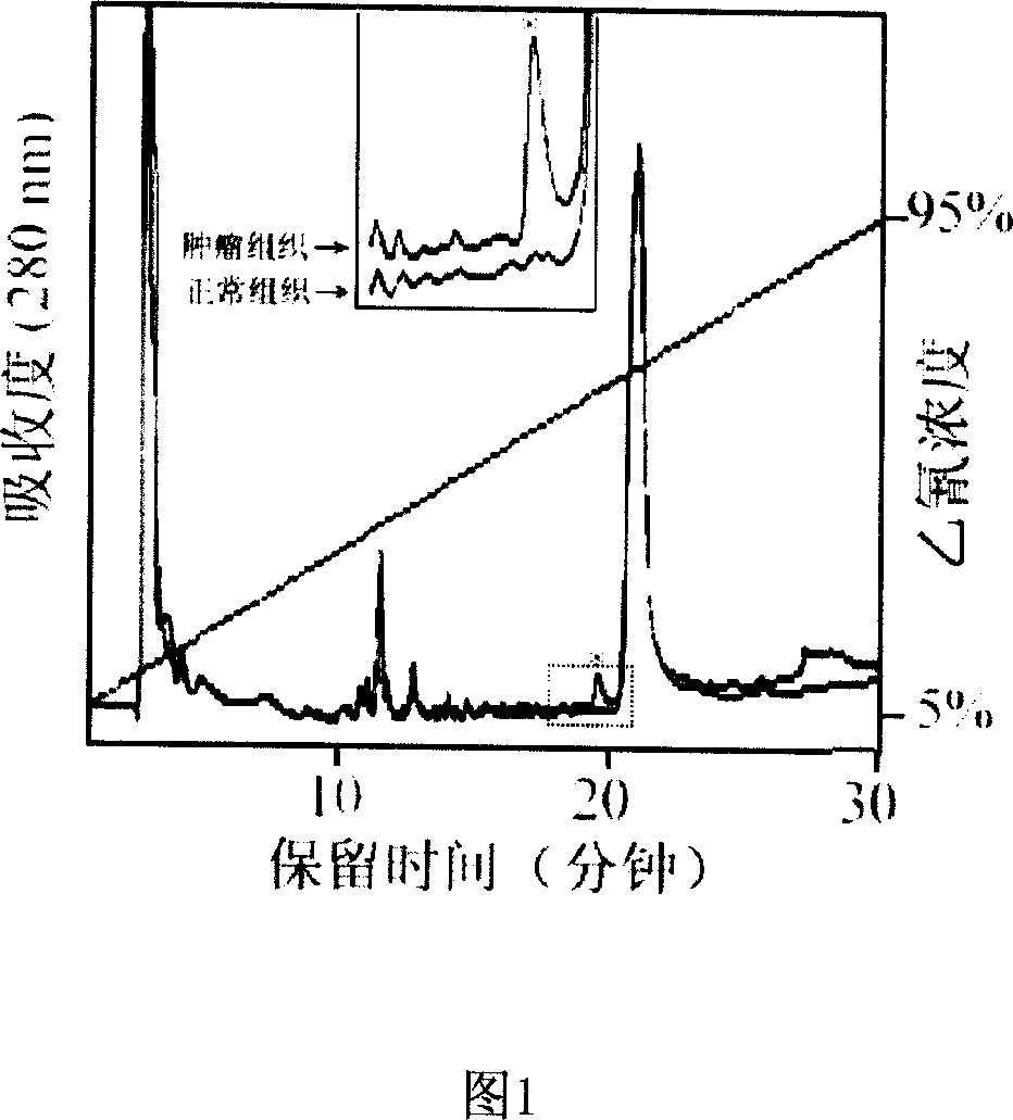
[0016] Example 1 High performance liquid chromatography (HPLC) method to detect the expression difference of HNP-1, -2, -3 protein in tumor tissue and its adjacent normal tissue
[0017] 1. Experimental Materials and Instruments
[0018] 15 pairs of colorectal cancer and its adjacent normal colorectal tissues, 5 pairs of colorectal cancer metastasized lung cancer tissues and their adjacent normal lung tissues, 15 pairs of gastric cancer tissues and their adjacent normal gastric tissues, the above tissues were obtained from the Second Affiliated Hospital of Zhejiang University Acquired from the Tissue Bank of the Cancer Institute. Analytical pure acetocyanide (CAN), acetic acid, trifluoroacetic acid (TFA), liquid chromatograph (HPLC) is the HEWLETT PACKARD HP series 1100 product of U.S. Hewlett Packard Company, and the separation column of HPLC is the C18 column (2.0 ×150mm, 300A).
[0019] 2. Experimental method
[0020] Take the tissue sample out of liquid nitrogen and was...
Embodiment 2
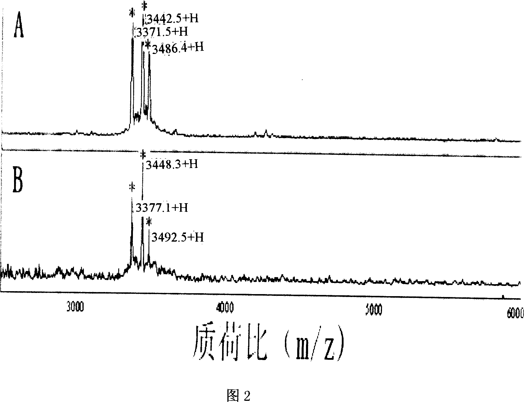
[0027] Example 2 Identification of NP-1, -2, -3 proteins by surface-enhanced laser desorption ionization-time-of-flight-mass spectrometry (SELDI-TOF-MS)
[0028] 1. Experimental materials
[0029] CHCA; gold chip; SELDI-TOF-MS mass spectrometer (CIPHERGEN, USA), DTT (dithiothreitol), NP20 chip of All-in-one standard protein.
[0030] 2. Experimental method
[0031] Collect the above-mentioned characteristic absorption peaks obtained by HPLC analysis, that is, the elution peak at the 63% elution concentration of CAN, dissolve the protein with an appropriate amount of tissue lysate (20% acetonitrile and 2% acetic acid) after freeze-drying, Take 1 μl of protein solution and apply it to the sample hole of the gold chip, add 0.5 μl of CHCA surface enhancer after drying at room temperature, perform SELDI-TOF-MS analysis after room temperature drying and use Ciphergen proteinchip 3.1 software to read the data. Before data collection, the SELDI instrument was calibrated by the All-i...
Embodiment 3
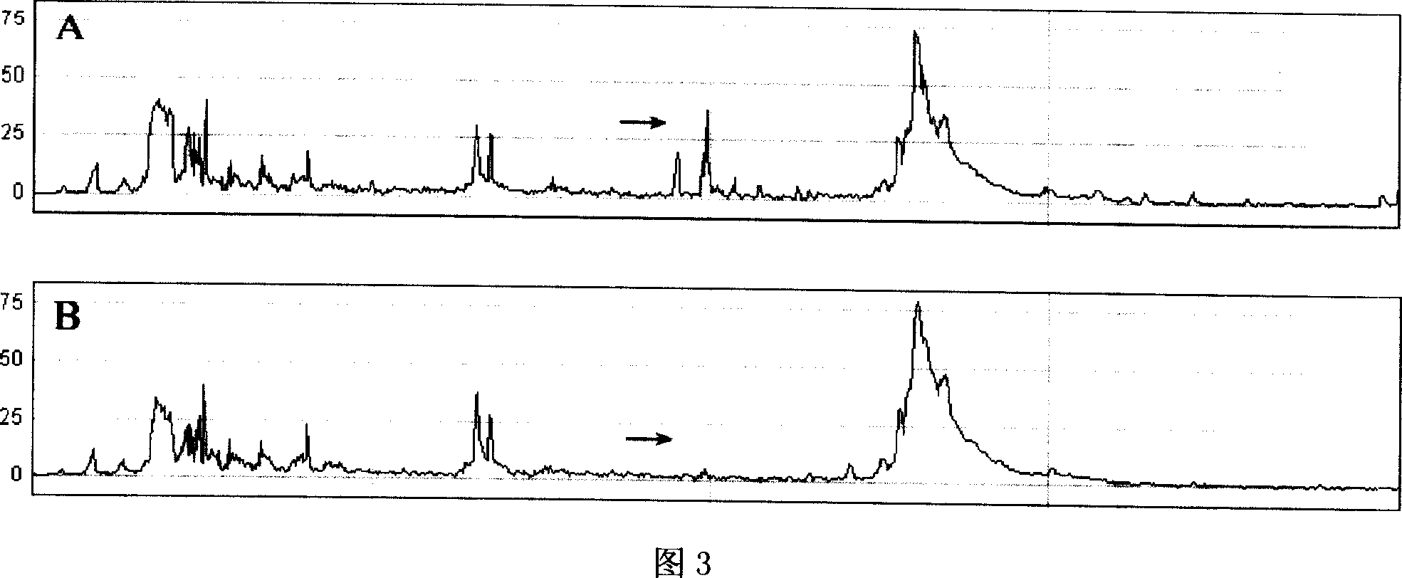
[0037] Example 3 Comparison of HNP-1, -2, -3 protein differences in tumor tissue and its adjacent normal tissue by flight mass spectrometry SELDI-TOF-MS
[0038] 1. Experimental materials
[0039] CHCA, gold chip, SELDI-TOF-MS mass spectrometer (CIPHERGEN, USA), DTT, NP20 chip of All-in-one standard protein.
[0040] 2. Experimental method
[0041] Take the tissue protein solution extracted in Example 1, and adjust the concentration of each tissue protein solution to 2 μg / μl. 1 μl of the above protein solution was taken from each tissue sample for SELDI analysis, and the SELD analysis and data collection methods were the same as in Example 2.
[0042] 3. Experimental results
[0043] See Figure 3 for the comparison results of SELDI analysis. Figure 3A is the SELDI-TOF-MS analysis chart of protein in lysate of lung cancer tissue with gastric cancer, colorectal cancer and colorectal cancer metastasis; Figure 3B is the protein expression of normal tissue lysate in adjacent ti...
PUM
 Abstract
Description
Claims
Application Information
 - R&D
- Intellectual Property
- Life Sciences
- Materials
- Tech Scout
- Unparalleled Data Quality
- Higher Quality Content
- 60% Fewer Hallucinations
Browse by: Latest US Patents, China's latest patents, Technical Efficacy Thesaurus, Application Domain, Technology Topic, Popular Technical Reports.
© 2025 PatSnap. All rights reserved.Legal|Privacy policy|Modern Slavery Act Transparency Statement|Sitemap|About US| Contact US: help@patsnap.com


