Systems and methods for quantitative analysis of histopathology images using multiclassifier ensemble schemes

a multi-classifier and ensemble scheme technology, applied in the field of cancer detection and classification, can solve the problems of variability in interpretation and application of grading criteria, inability to reproduce judgments, and difficulty in quantitative analysis and classification

Active Publication Date: 2018-08-21
THE BOARD OF TRUSTEES OF THE LELAND STANFORD JUNIOR UNIV +1
View PDF30 Cites 15 Cited by
  • Summary
  • Abstract
  • Description
  • Claims
  • Application Information

AI Technical Summary

Benefits of technology

[0029]One embodiment of the present invention introduces a variable-rate learning algorithm for quaternion neural networks. The learning rate is varied at each training cycle according to the maximum Mean Square Error (MSE) computed for any single training pattern over all of the neuron outputs, though other heuristics besides MSE may also be employed. This learning rate adjustment algorithm lowers the learning rate as the network comes closer to convergence, in order to avoid significant problems with misadjustment.
[0032]The exemplary computer-aided diagnosis systems of the present disclosure are robust in terms of accuracy, sensitivity, specificity, positive predictive value, and negative predictive value; and may be used to achieve standardized and reproducible cancer classification outcomes.

Problems solved by technology

These judgments may not be reproducible due factors including the pathologist's skill level, fatigue, and experience.
Moreover, there may be variability in the interpretation and application of the grading criteria, particularly when grading or scoring complicated tissue samples.
The inherent complexity and non-homogeneity of histopathology images make quantitative analysis and classification a challenging task.
However, using this method, correlations between color channels are lost.
Since humans perceive thousands of colors but only ten to fifteen levels of grayscale, present classification methods do not necessarily provide optimal performance.
However, real-valued neural networks process images in monochrome, therefore leading to the aforementioned problems with recombining the color planes.
A major drawback to quaternion neural networks is their computational cost: The multiplication of two quaternions requires sixteen real multiplications, instead of one multiplication for real numbers.
A significant drawback of this method is that additional an NIR stain must be used; the most widespread in clinical practice is H&E stain.
A drawback of the system is that it is not able to grade or score a detected cancer region.
Unfortunately, while this level of automation has improved the physical handling of pathology images, it has provided no concrete assistance to pathologists such that the accuracy of diagnosis needs to be improved [WO 2009006696 A1].

Method used


Image

  • Systems and methods for quantitative analysis of histopathology images using multiclassifier ensemble schemes
  • Systems and methods for quantitative analysis of histopathology images using multiclassifier ensemble schemes
  • Systems and methods for quantitative analysis of histopathology images using multiclassifier ensemble schemes

Examples


example 1

Gleason Grading

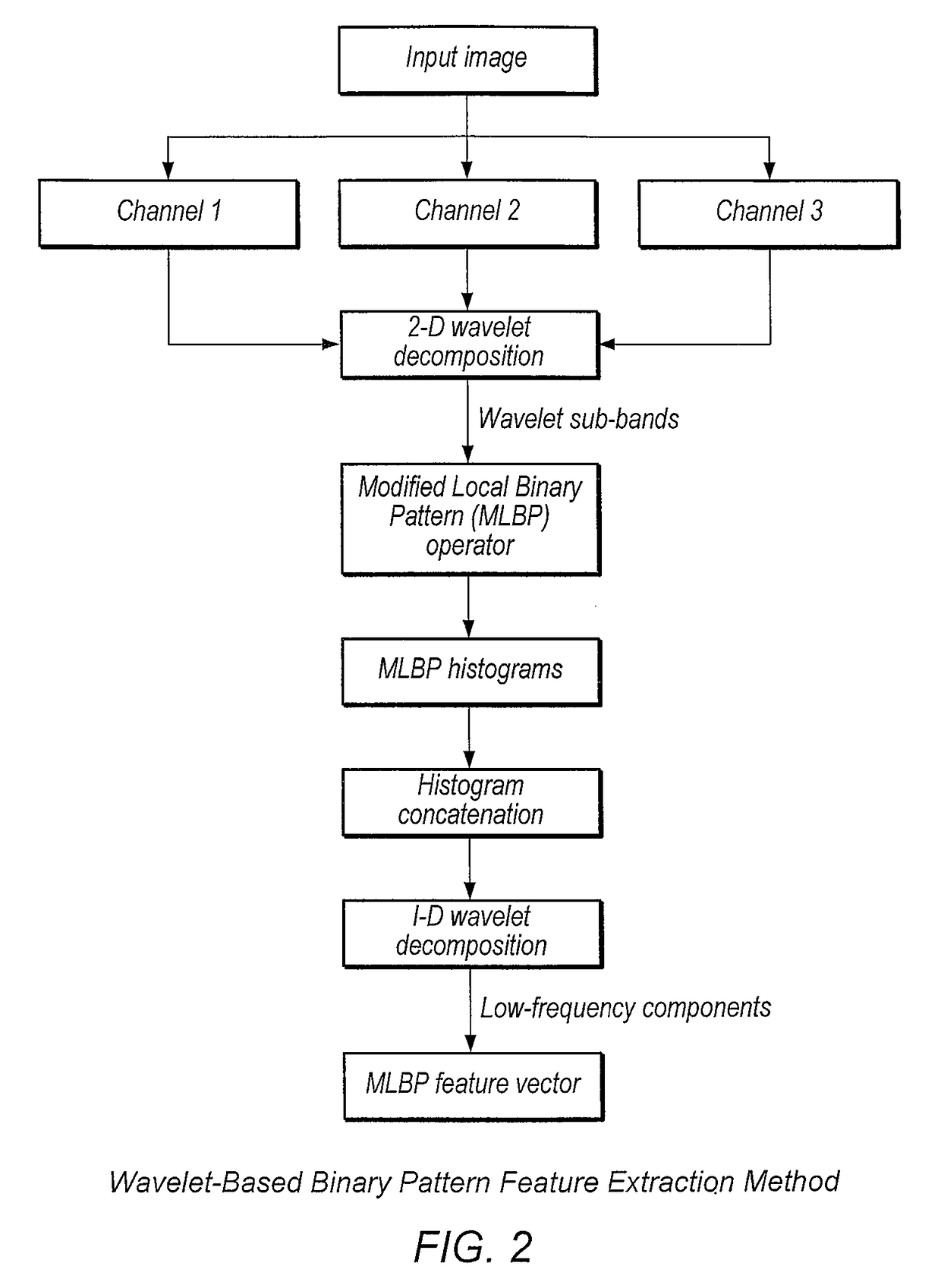
[0123]In this example a prostate cancer Gleason grading system was used to estimate the prostate cancer grade. Described herein is the framework for the development of a prostate cancer grading system. For purposes of the illustration of this embodiment of the present invention an example of a system for the grading of prostatic carcinoma from digitized histopathology slides is explained in detail. However, it must be understood that the present invention is also applicable to biopsies of other body organs including but not constrained to the skin, liver, breast, brain, kidney, pancreas, lungs, colon, rectum, bladder, cervix, and uterus. The input data may be small images of pre-selected regions of interest or whole-slides. When processing whole-slides, a grid of size s×s is placed over the image, and the classification process described below is performed once per resulting block. The general architecture of the system is illustrated in FIG. 5.

[0124]The description o...

example 2

Prostate Cancer

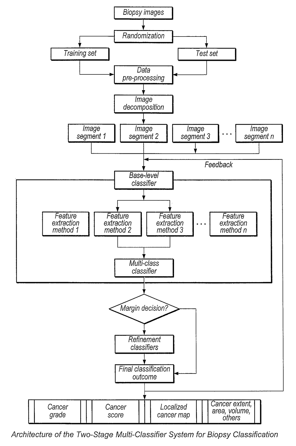
[0175]In this example a multi-classifier system was used to grade prostate cancer. Described herein is a system for grading and scoring of prostate cancer based on the Gleason grading system, which is presented as an exemplary application of the multi-classifier system for biopsy classification. The system is composed of a pre-processing unit and six base-level sub-systems. Each base-level sub-system is comprised of a feature extraction unit, a classification method, and, as necessary, a function to obtain posterior probabilities from the classification output. In addition, the multi-classifier system has a fusion unit and several binary classifiers for refinement tasks. The decision of the classifier ensemble is produced by using a dynamic weighted voting technique, which will be explained later in this section. The purpose of the binary refinement classifiers is to improve overall system accuracy.

[0176]The description of processes carried out in the system will be s...

example 3

Skin Cancer Classification

[0223]In this example a Multi-classifier system was used to detect skin cancer. In order to demonstrate the general applicability of the disclosed system, the multi-classifier system of the previous example is also applied to the problem of skin cancer classification. The output of the system provides information about the nature of the lesion by classifying input data into major types of skin cancers: melanoma (MM), basal cell carcinoma (BCC), and cutaneous squamous cell carcinoma (SCC). Experiments were carried out on a database of 60 color images of Hematoxylin and Eosin (H&E)-stained skin tissue samples. The dataset contains 30 images of melanoma, 20 images of BCC, and 10 of SCC. Leave-one-out cross-validation was used to estimate the performance of the system. The average CCR, sensitivity, specificity, PPV, and NPV are summarized in Table 3.

[0224]

TABLE 3Performance indicators of the proposed system in skin cancer classification averaged over 10 simulat...


PUM


Abstract

Described herein are systems and methods for performing multi-stage detection and classification of cancer regions from digitized images of biopsy slides. Novel methods for processing the digitized images to improve feature extraction and structure identification are disclosed, including but not limited to the use of quaternions, logarithmic mappings of color channels, and application of wavelets to logarithmic color channel mappings. The extracted features are utilized in improved machine learning algorithms that are further optimized to analyze multiple color channels in multiple dimensions. The improved machine learning algorithms include techniques for accelerating the training of the algorithms, making their application to biopsy detection and classification practical for the first time. The performance of the described systems and methods are further improved by the disclosure of a novel multistage machine learning scheme, in which additional classifiers are utilized to choose among the classes proposed by other classifiers in close cases.

Description

BACKGROUND OF THE INVENTION[0001]1. Field of the Invention[0002]The invention generally relates to cancer detection and classification. The present disclosure includes quantitative analysis of and predictive modeling from digitized histopathology images. This disclosure includes methods that integrate multiple image description methods and classification methods into robust computer systems for accurate detection and classification or grading of cancerous tissue samples.[0003]2. Description of the Relevant Art[0004]The microscopic analysis of histopathology samples is widely used as a cancer diagnosis method. Histopathology is a labor-intensive process requiring the preparation of biopsy material including fixation, freezing, sectioning and staining followed by analysis and interpretation. Together these process steps have traditionally led to turnaround times of days rather than hours. Furthermore, tissue assessment is routinely performed by human visual evaluation of the structura...

Claims


Application Information

Patent Timeline
Patent Type & Authority Patents(United States)
IPC IPC(8): G06K9/00G16H50/20G06F19/00G06N5/04G06N99/00G06K9/62G06T7/00G06K9/34G06K9/46G06K9/66G06T7/246G06N3/04G06N20/20G16Z99/00
CPCG16H50/20G06K9/34G06K9/4652G06K9/628G06K9/629G06K9/6212G06K9/66G06N3/0427G06N5/043G06N99/005G06T7/0012G06T7/0014G06T7/246G06K9/0014G06T2207/30096G06K2009/4666G06T2207/10024G06T2207/10056G06T2207/20036G06T2207/20064G06T2207/20081G06T2207/20084G06T2207/30024G06N20/00G06N20/20G16Z99/00G06V20/695G06V10/467G06V10/758G06V10/806G06N3/042G06F18/253G06F18/2431
Inventor AGAIAN, SOSMOSQUERA-LOPEZ, CLARA M.GREENBLATT, AARON
Owner THE BOARD OF TRUSTEES OF THE LELAND STANFORD JUNIOR UNIV
Why Eureka
Try Eureka