System and method for ultrafast magnetic resonance spectroscopic imaging using learned spectral features

a magnetic resonance spectroscopic and learning technology, applied in the field of magnetic resonance spectroscopic imaging, can solve the problems of slow development of research and clinical applications, poor linear fitting method, and inability to provide the signal-to-noise ratio (snr), resolution and speed, and the current mrsi method is still far behind in providing the signal-to-noise ratio (snr)

Active Publication Date: 2021-08-03
THE BOARD OF TRUSTEES OF THE UNIV OF ILLINOIS
View PDF15 Cites 0 Cited by
  • Summary
  • Abstract
  • Description
  • Claims
  • Application Information

AI Technical Summary

Problems solved by technology

The linear fitting method yielded poor fitting in regions of large field inhomogeneity (highlighted by red arrows in middle and rightmost columns of portion (b) of FIG. 10) when a large number of echoes were used.
However, even after four decades of development, the existing MRSI methods are still far behind in providing the signal-to-noise ratio (SNR), resolution and speed desired for practical applications.
The unique power of MRSI as a label-free molecular imaging modality has long been recognized 4A, 15A, but its research and clinical applications have been developing slowly due to a number of fundamental technical hurdles.
Specifically, MRSI adds one or more spectral dimensions to the image function of interest, making the imaging problem higher dimensional.
Accordingly, the number of spatiospectral encodings required in the conventional Nyquist sampling paradigm grows exponentially as the resolution increases, which leads to the slow imaging speed.
However, even with all these efforts, state-of-the-art MRSI methods are still limited to a centimeter-level resolution, around half an hour imaging time, and very limited organ coverage 37A.
Furthermore, for 1H-MRSI in particular, effective separating the three to four orders of magnitude stronger water and lipid signals from the metabolite signals of interest is also a long-standing challenge.
A common practice to address this is to incorporate special water and lipid signal suppression modules into the MRSI pulse sequences 38A-40A, which strongly limit the acquisition design yet complete suppression is impossible.
Furthermore, each of these encodings is required to have high SNR due to the low molecular concentrations of the metabolites of interest embedded in ρ(r, f), thus the very slow imaging speed and low resolution (large voxel size) commonly observed in MRSI experiments.
However, due to the requirement of high-SNR data (56A, 57A), the applications of CS to MRSI have been limited, e.g., to accelerate hyperpolarized (13A) C-MRSI (58A) or very low-resolution acquisitions (59A-61A).
However, such undersampling alone is still limited to the inherent SNR efficiency of SE-based acquisition and does not fully take advantage of the power of the subspace model in reducing spectral sampling (i.e., still requiring longer TRs to maintain SNR efficiency (65A)).
Such a special acquisition strategy imposes unique challenges for spatiospectral processing.
With such a model, the key issues are the determination of the subspaces (i.e., {ϕlx(f)}lx=1Lx) and the use of these subspace for effective separation of different signal components and high-SNR metabolite reconstruction from noisy data.
Furthermore, because of the extended k-space coverage along kx to achieve the desired resolution, the spacing between gradient echoes does not satisfy the spectral Nyquist (16A, 17A).
Currently, QSM and MRSI are carried out in separate scans, both of which often require long data acquisition times, limiting their practical use in clinical and research applications.
There are three key data processing problems in deriving the susceptibility and metabolite maps from the measured data: (1) reconstruction of the spatiospectral / spatiotemporal function from the sparsely sampled (k,t)-space data, (2) estimation of the susceptibility maps from the reconstructed spectroscopic signals, and (3) reconstruction of the metabolite signals.
However, a full analysis of its performance is needed, which is beyond the scope of the discussion herein.
Second, SPICE has a relatively smaller k-space coverage than conventional QSM methods.
Note also that the linear phase method yielded some “bad” fitting points (red arrows in portion (b) of FIG. 10) in regions with large field inhomogeneity, especially when a large number of echoes was used (because of unreliable phase unwrapping for noisy data at long echo times).
Note that because of the multiple nonlinear processing steps involved, a full characterization of the SNR and resolution of the final QSM maps is challenging.
However, the methods described herein do not prevent the use of sensitivity determined from auxiliary high-resolution anatomical scans as long as data registration is properly done.

Method used


Image

  • System and method for ultrafast magnetic resonance spectroscopic imaging using learned spectral features
  • System and method for ultrafast magnetic resonance spectroscopic imaging using learned spectral features
  • System and method for ultrafast magnetic resonance spectroscopic imaging using learned spectral features

Examples


Embodiment Construction

[0035]Embodiments are directed toward a method and apparatus to enable ultrafast, high-resolution MRSI.

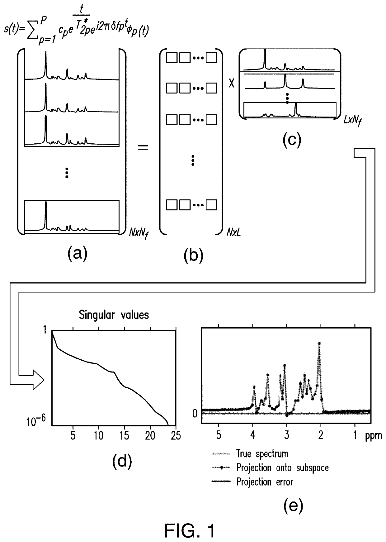
[0036]In one or more embodiments, spectral features of MR-detectable molecules (e.g., N-Acetylaspartate, Creatine, Choline, Glutamate, Glutamine, Myo-inositol, Glutathione, GABA, Lactate, Taurine, Aspartate in 1H-MRSI experiments or Phosphocreatine, ATPs, Inorganic phosphate, NADH, Phosphomonoesters, Phosphodiesters in 31P MRSI experiments) are predetermined (or learned) and used to calculate the spatiospectral distributions from measured MRSI data.

[0037]In one or more embodiments, spectral features of MR-detectable molecules will be learned from a set of “training” data acquired a priori, taking into account the resonance structure of each compound, which can be predetermined based on MR physics (using quantum simulation, for example) and the data acquisition scheme used.

[0038]In one or more embodiments, “training” data for metabolites can be obtained using, single-voxel (SV) scan...


PUM

PropertyMeasurementUnit
voxel sizeaaaaaaaaaa
static magnetic fieldaaaaaaaaaa
magnetic fieldaaaaaaaaaa

Abstract

A new method is developed for ultrafast, high-resolution magnetic resonance spectroscopic imaging (MRSI) using learned spectral features. The method uses Free Induction Decay (FID) based ultrashort-TE and short-TR acquisition without any solvent suppression pulses to generate the desired spatiospectral encodings. The spectral features for the desired molecules are learned from specifically designed “training” data by taking into account the resonance structure of each compound generated by quantum mechanical simulations. A union-of-subspaces model that incorporates the learned spectral features is used to effectively separate the unsuppressed water/lipid signals, the metabolite signals, and the macromolecule signals. The unsuppressed water spectroscopic signals in the data can be used for various purposes, e.g., removing the need of additional auxiliary scans for calibration, and for generating high quality quantitative tissue susceptiability mapping etc. Simultaneous spatiospectral reconstructions of water, lipids, metabolite and macromolecule can be obtained using a single 1H-MRSI scan.

Description

CROSS-REFERENCE TO RELATED APPLICATION(S)[0001]The present application is a U.S. National Stage of International Application No. PCT / US2018 / 047774, having an International Filing Date of Aug. 23, 2018, which claims the benefit of priority to U.S. Provisional Patent Application No. 62 / 552,193, filed on Aug. 30, 2017, the disclosure of which is incorporated by reference herein in its entirety (including each appendix, if any, thereof).[0002]International Application No. PCT / US2018 / 047774 (of which the present application is a U.S. National Stage) is related to U.S. patent application Ser. No. 14 / 992,498, filed Jan. 11, 2016 (published as U.S. Patent Application Publication No. 2016 / 0202336), the disclosure of which is incorporated by reference herein in its entirety (including each appendix, if any, thereof).STATEMENT AS TO FEDERALLY SPONSORED RESEARCH[0003]This invention was made with government support under contract number R21-EB021013 awarded by the National Institutes of Health. ...

Claims


Application Information

Patent Timeline
Patent Type & Authority Patents(United States)
IPC IPC(8): G01R33/485A61B5/00A61B5/055G01R33/24G01R33/34G01R33/46G01R33/465G01R33/48G01R33/56G01R33/561
CPCG01R33/485A61B5/0042A61B5/055G01R33/246G01R33/34092G01R33/465G01R33/4625G01R33/4816G01R33/4826G01R33/4828G01R33/5608G01R33/5615A61B5/7253G01R33/5616
Inventor LIANG, ZHI-PEILAM, FAN
Owner THE BOARD OF TRUSTEES OF THE UNIV OF ILLINOIS
Why Eureka
Try Eureka