Use of pro-apoptotic factors in treatment of atherosclerosis

a pro-apoptotic factor and atherosclerosis technology, applied in the field of vascular disease, can solve the problems of reduced lumen size and tissue perfusion, limited blood flow through the vessel, and treatment does not achieve an acute reduction in the size of the vascular lesion

Inactive Publication Date: 2001-12-20
BETH ISRAEL DEACONESS MEDICAL CENT INC
View PDF0 Cites 2 Cited by
  • Summary
  • Abstract
  • Description
  • Claims
  • Application Information

AI Technical Summary

Problems solved by technology

The fundamental pathology of vascular disease is an abnormal accumulation of cells within the subintimal space below the surface of the endothelial cell lining, resulting in a decrease in lumen size and tissue perfusion.
This accumulation is due to the proliferation and / or migration of smooth muscle cells, and / or inflammatory cells into the intimal layer of a vessel, resulting in restricted blood flow through that vessel, i.e. neointimal occlusive lesions.
Despite the substantial benefit attributable to the use of current cholesterol lowering and immunosuppressive drug therapies, these treatments do not achieve an acute reduction in vascular lesion size.
Furthermore, bypass surgery may accelerate progressive lesion stenosis, and interventions such as balloon angioplasty often result in the development of restenosis.
EC activation promotes inflammation and can lead to apoptosis of these cells.
Clinical trials aimed at blocking a single growth or inflammatory factor have failed to show efficacy.

Method used


Image

  • Use of pro-apoptotic factors in treatment of atherosclerosis
  • Use of pro-apoptotic factors in treatment of atherosclerosis
  • Use of pro-apoptotic factors in treatment of atherosclerosis

Examples


example 1

A20 Activity in SMC

[0121] A20 is Part of the Physiological response of SMC to TNF

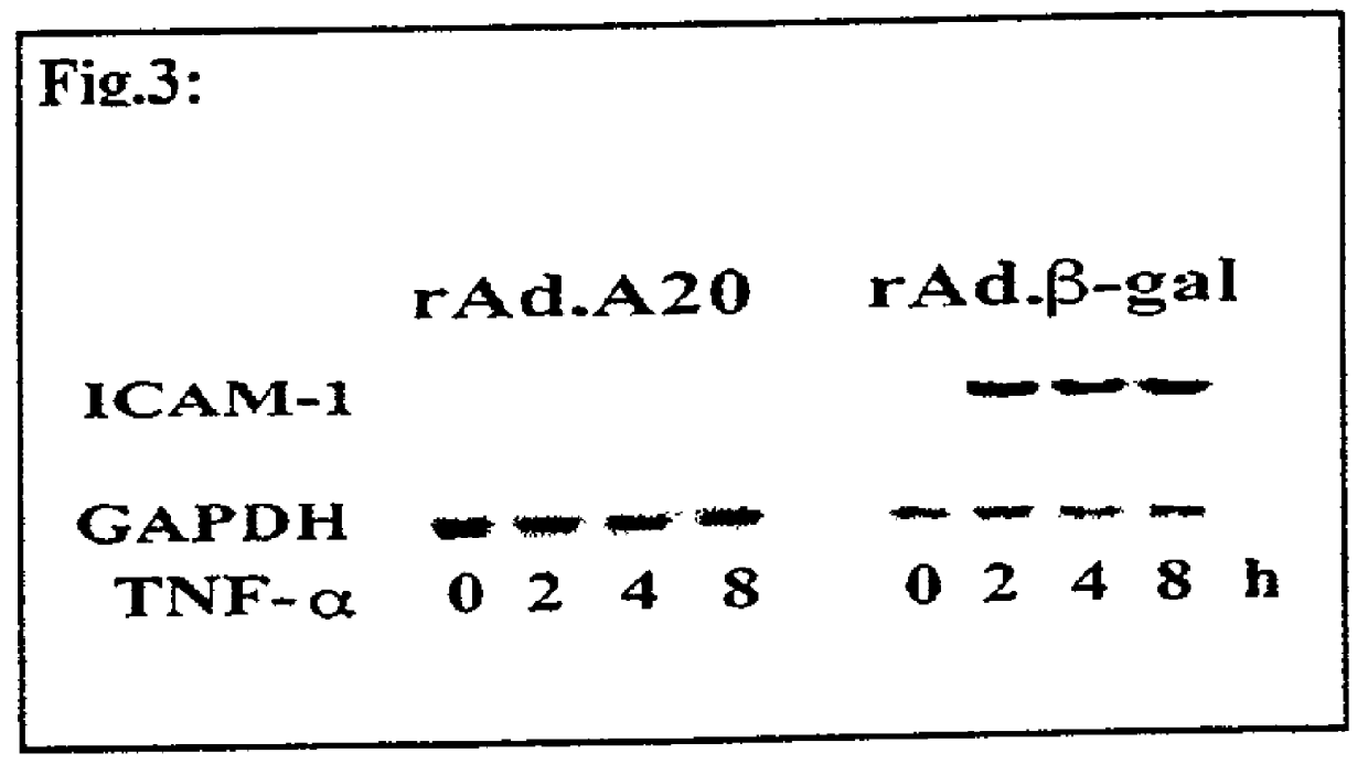
[0122] Primary human aortic SMC were cultured in 6 well plates using smooth muscle basal medium (SmBM) (Clonetics, California) supplemented with growth factors, Gentamycin and Amphotericin B. Confluent cells were stimulated with 100 Units of recombinant human (rhu)TNF and RNA was extracted before and at 1 and 6 hours (h) following addition of TNE. RNA samples were analyzed by Northern blot analysis for the expression of A20 using an A20 cDNA probe (Ferran et al., supra). In all experiments, a cDNA probe for human GAPDH was used to evaluate equal loading of RNA. Results showed almost no A20 mRNA expression prior to TNF addition. A20 mRNA was strongly induced 1 h following TNF stimulation and started declining 6 h thereafter (FIG. 1A). The induction of A20 by TNF was confirmed at the protein level by means of immunohistochemistry using a rabbit anti-human A20 polyclonal antibody developed in the laborator...

example 2

Evaluation of the Effects of Expressing A20 in SMC In Vitro on Inhibition of NF-.kappa.B Activation and the Impact on SMC Activation and Proliferation Induction of A20

[0143] One may determine whether A20 expression is part of the physiological response of the SMC to injury.

[0144] Fourth to sixth passage human aortic SMC monolayer cultures are activated using different agonists that are relevant to the pathogenesis of atherosclerosis. These agonists include TNF, LDL, and oxidized LDL, growth factors (platelet derived growth factor (PDGF)) and CD40 cross-linking. The expression of A20 in SMC following these agonists is evaluated at different time points (1-24 h), both at the mRNA and protein levels as described in the preliminary results. One then tests whether each of these critical mediators of the atherosclerotic lesion will, like TNF, induce the expression of A20. We propose, without limiting the biochemical mechanism of the invention, that A20 induction is part of the "regulatory...

example 3

A20 Activity In Vivo and In Vitro

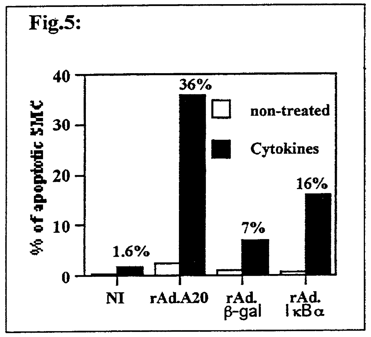
[0154] Impact of A20 Overexpression Upon SMC Apoptosis In Vitro

[0155] Apoptosis is now viewed as beneficial to prevent redevelopment and promote the regression of established atherosclerotic lesions. The present invention demonstrates for the first time that A20 sensitizes SMC to cytokine-mediated apoptosis, making A20 a prime gene therapy target to achieve this aim. These experiments are planned to extend our finding that A20 sensitizes SMC to cytokine-mediated apoptosis and to other apoptotic stimuli that are present within the atherosclerotic plaque i.e. Fas, NO and oxidized LDL. Second, one may determine the molecular basis of the effect of A20 upon the death signaling machinery. These studies evaluate the effect of A20 on activation of caspases, mitochondrial membrane potential, c-myc, cytochrome c release and cleavage of death substrates such as PARP that are the hallmarks of apoptosis.

[0156] As described above, non-infected SMC and SMC infecte...


PUM

PropertyMeasurementUnit
timeaaaaaaaaaa
temperatureaaaaaaaaaa
pHaaaaaaaaaa

Abstract

The present invention features a novel method of treating vascular disease that involves modifying smooth muscle cells to express a gene encoding a protein having both anti-inflammatory and pro-apoptotic activity. Preferably, the protein of the invention also has an anti-proliferative effect in smooth muscle cells. In general, the method is useful in preparing vascularized organs and vessels for transplant into a patient. Alternatively, the present invention can be applied to treat atherosclerotic lesions in damaged vessels.

Description

[0001] The present application claims benefit to Provisional application No. 60 / 177,535 filed Jan. 21, 2000, the entire contents of which are incorporated herein by reference.Statement as to Federally Sponsored Research[0002] The present research was supported by a grant from the National Institutes of Health (NIH) (Grant R01 Number HL57791-02).[0003] 1. Field of the Invention[0004] The present invention relates to the field of vascular disease.[0005] 2. Background of the Invention[0006] Vascular disease is the most common cause of morbidity and mortality in the western world, surpassing any other single degenerative disease. The fundamental pathology of vascular disease is an abnormal accumulation of cells within the subintimal space below the surface of the endothelial cell lining, resulting in a decrease in lumen size and tissue perfusion. This accumulation is due to the proliferation and / or migration of smooth muscle cells, and / or inflammatory cells into the intimal layer of a v...

Claims


Application Information

Patent Timeline
Patent Type & Authority Applications(United States)
IPC IPC(8): A61K35/12A61K35/34A61K38/17A61K38/19A61K48/00C12N5/071C12N5/077
CPCA61K38/1709A61K38/177A61K38/19A61K48/00C12N5/0661C12N5/0691C12N2503/02C12N2510/00A61K35/34A61K2300/00
Inventor FERRAN, CHRISTIANEARVELO, MARIA B.
Owner BETH ISRAEL DEACONESS MEDICAL CENT INC
Why Eureka
Try Eureka