

Method for culturing cells
a cell culture and cell technology, applied in the field of cell culture methods, can solve the problems of inability to isolate and culture microvascular endothelial cells, inability to isolate substantially purified populations of microvascular endothelial cells from any tissue source, and inability to achieve cell culture successfully, etc., to achieve the effect of enhancing the growth of microvascular endothelial cells
- Summary
- Abstract
- Description
- Claims
- Application Information
AI Technical Summary
Benefits of technology
Problems solved by technology
Method used
Image

Examples
example 2
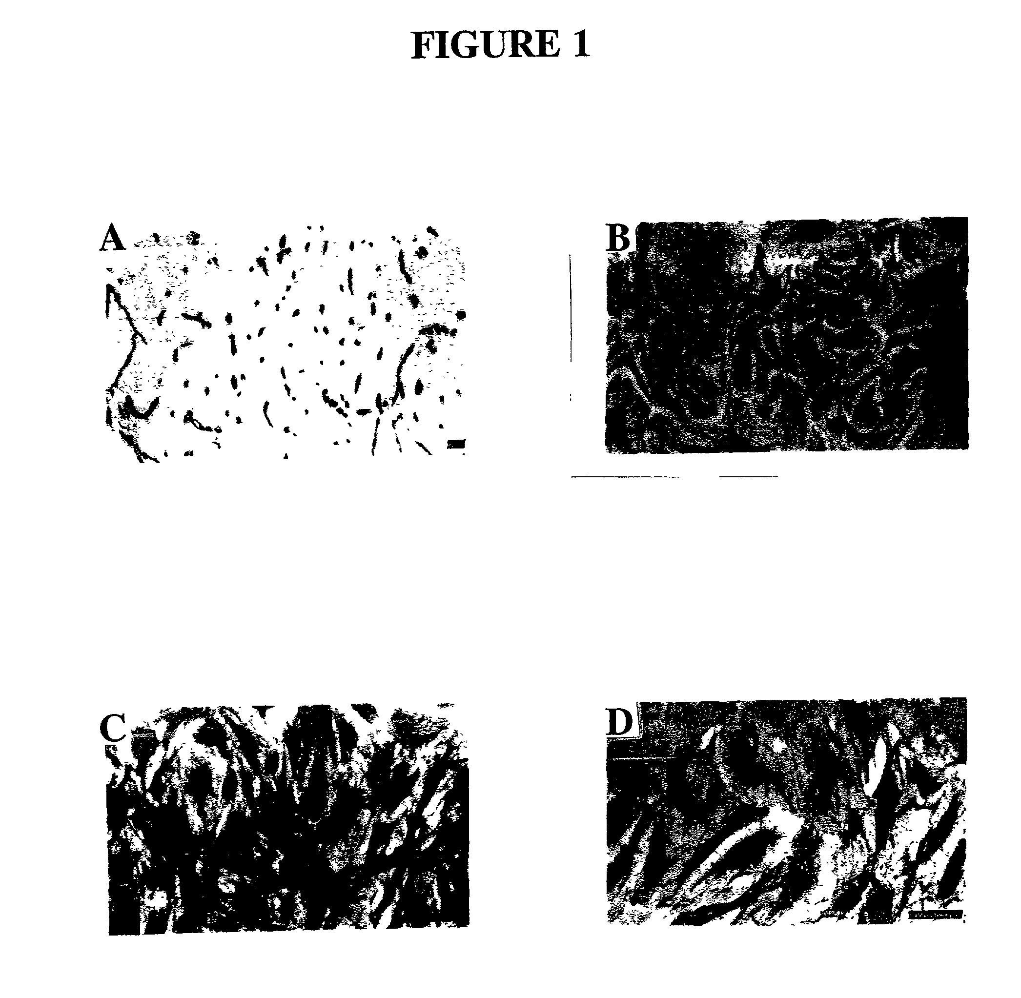
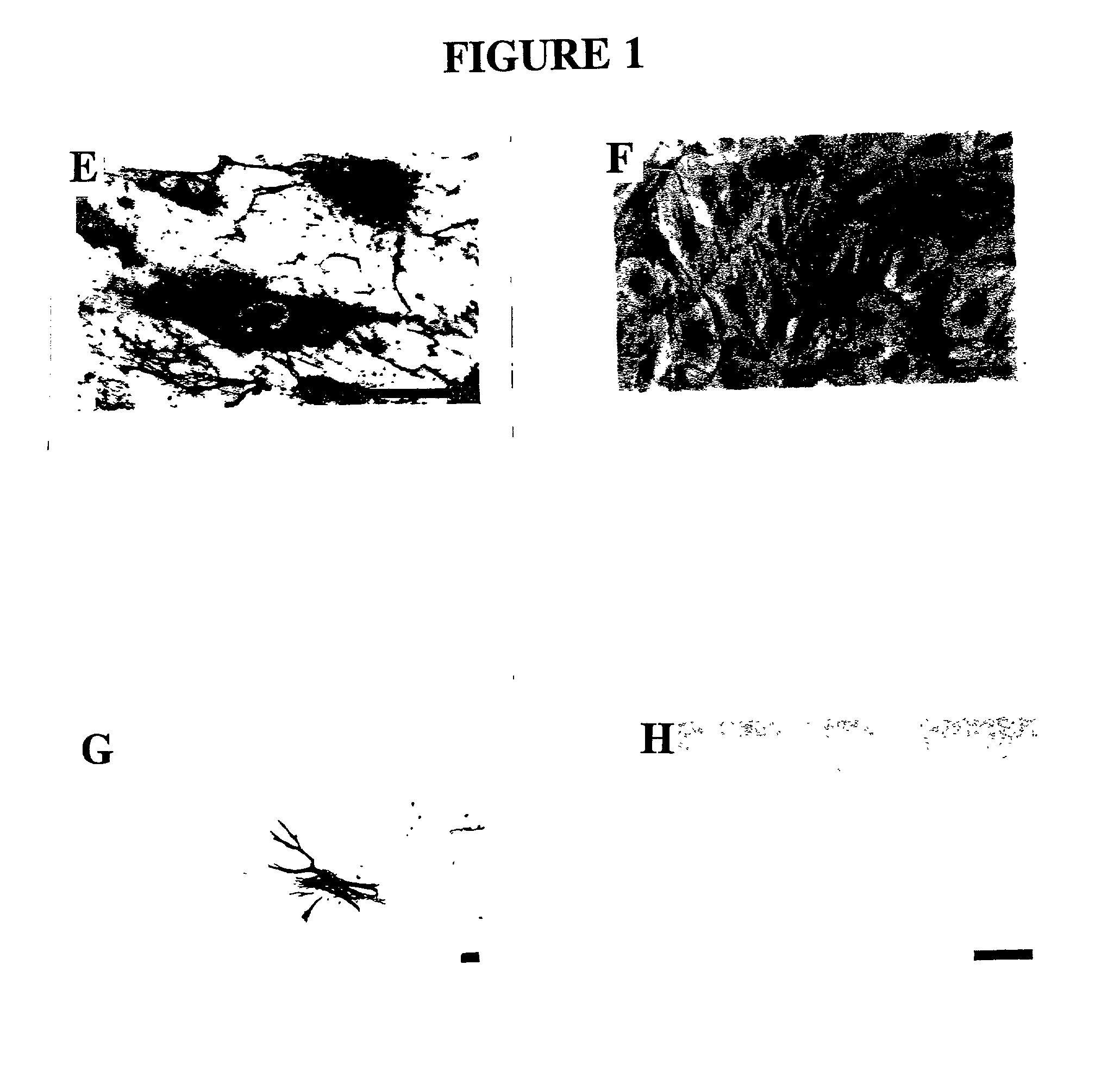
Microvascular Endothelial Cell Isolation and Purification
[0135] The endometrial layer of the uterine tissue was identified, removed and discarded together with the first 1 mm of myometrial tissue and the outer third of the myometrium, thus removing any of the serosa and mesothelial layer. The inner 2 / 3 of the myometrium was then finely chopped for enzyme dissociation, since the inner layers of myometrium show greater responses to sex steroid hormones compared to the outer third (Noe et al., 1999). Chopped tissue was digested with 2 mg / ml collagenase type 2 (Worthington, Biochemical Corporation, Freehold, N.J., USA) and 14.5 .mu.g / ml deoxyribonuclease type I (Boehringer Mannheim GmbH, Mannheim, Germany) in CA.sup.2+- and Mg.sup.2+-free phosphate buffered saline (PBS, pH 7.4) containing 0.1% BSA (Sigma Chemical Co., St. Louis, Mo., USA) for 2 hour at 37.degree. C. in a shaking water bath. The cloudy supernatant containing single cells was sequentially removed at 30 minute intervals du...
example 3
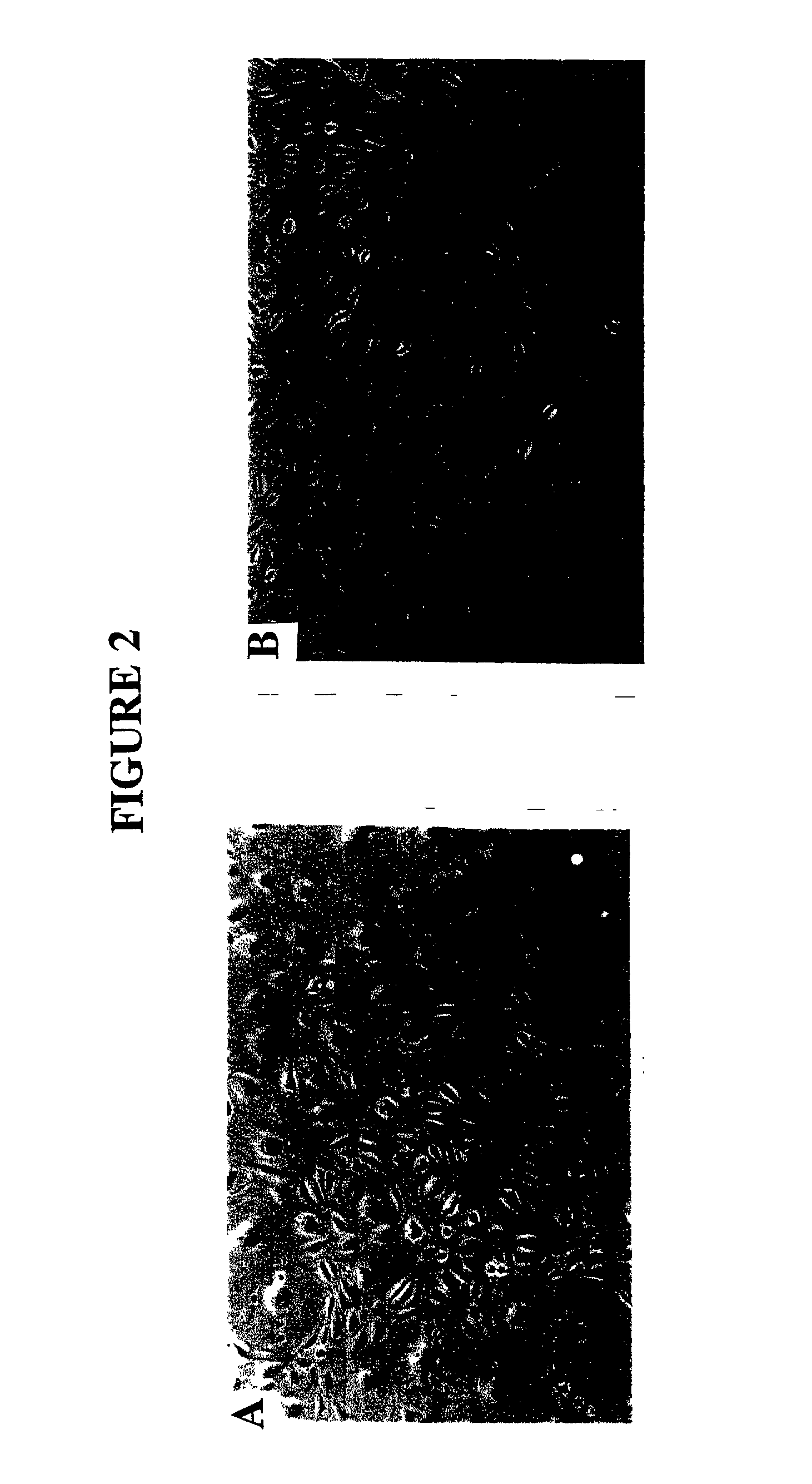
Microvascular Endothelial Cell Culture
[0138] The purified MEC were resuspended in a standard culture medium comprising M199 with Earle's salts containing heat-inactivated 15% male human serum (HS) (obtained from Red Cross Blood Service and male staff volunteers) and 5% FCS (CSL, Melbourne, Australia), 2 mM glutamine (Gibco BPL), 5 ng / ml bFGF (Gibco BRL), 0.1 mg / ml heparin (Gibco BRL), and antibiotic / antimycotic, seeded into culture flasks at 8-10.times.10.sup.4 cells / cm.sup.2 coated with 10 .mu.g / ml fibronectin (Gibco BRL) and incubated in a humidified atmosphere at 37.degree. C. in 5% CO.sub.2 in air. For some isolations 0.5 .mu.g / ml hydrocortisone (Sigma), 330 .mu.M 3-isobutyl-1-methyl xanthine (IBMX: Sigma), 2 mM MgSO.sup.4 (complex culture medium) were also included in the culture medium. Medium was changed every 2-3 days and at 70-80% confluence, MEC were trsinised (0.025% trypsin: 0.27 mM EDTA) and repurified with UEA-1 coated dynabeads and subcultured at a split ratio of 1:3 ...
example 4
Immunhistochemistry
[0139] MEC were grown on 13 mm gelatin-coated Thermanox coverslips (Nunc Roskilde, Denmark) and when confluent were rinsed in PBS and fixed in cold acetone for 2 minutes. Standard immunohistochemistry protocols (Abberton et al. 1999; Goodger (MacPherson) and Rogers, 1994; Gargett et al. 1999) were used after first blocking sections with 0.03% H.sub.2O.sub.2 for 10 minutes at room temperature (RT) and protein blocking reagent (PBA) (Lipshaw Immunon, Pittsburgh, Pa., USA) for 10 minutes at RT. The primary antibodies were then incubated for 1 hour at 37.degree. C., followed by biotinylated rabbit anti-mouse or goat anti-rabbit secondary antibodies (1 / 100) for 30 minutes at RT, streptavidin-HRP conjugate (Zymed, San Francisco, Calif., USA) for 30 minutes at RT and AEC chromagen (Zymed) for 10 minutes. The following primary antibodies were used: rabbit anti-human Factor VIII related antigen at 20 .mu.g / ml (Zymed), mouse anti-human CD31, 8.2 .mu.g / mnl (Zymed), mouse ant...
PUM
 Abstract
Description
Claims
Application Information