

Calcium phosphate particles as mucosal adjuvants
- Summary
- Abstract
- Description
- Claims
- Application Information
AI Technical Summary
Problems solved by technology
Method used
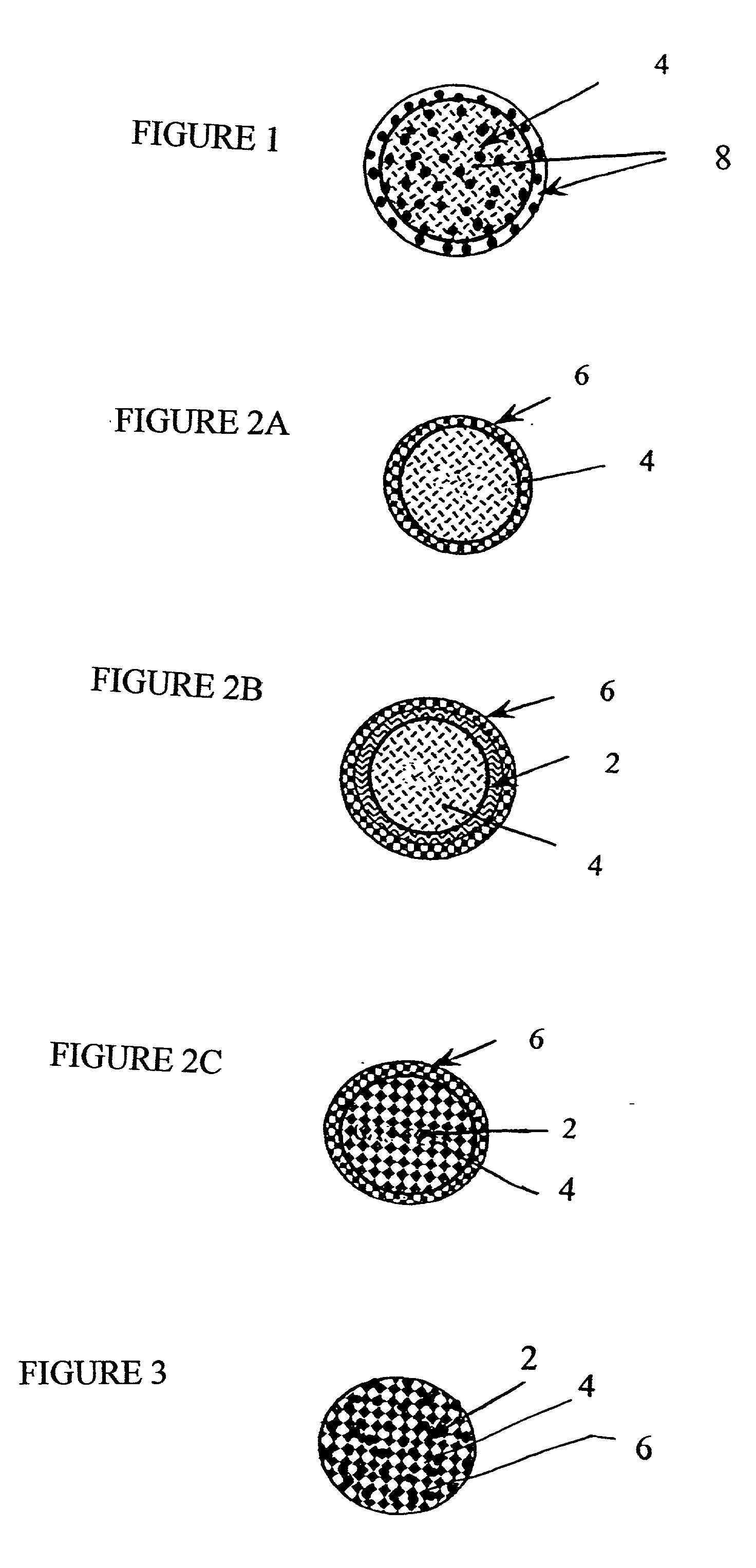
Image

Examples
example 1
[0115] A 12.5 mM solution of CaCl.sub.2 is prepared by mixing 1.8378 g of CaCl.sub.2 into 800 mL of sterile GDP water under aseptic conditions until completely dissolved, and the solution diluted to 1 L and filtered. A 15.625 mM solution of sodium citrate was prepared by dissolving 0.919 g of sodium citrate into 200 mL of sterile GDP water with mixing using aseptic techniques and filtered. A 12.5 mM solution of dibasic sodium phosphate was prepared by dissolving 1.775 g sodium phosphate into 1 L of sterile GDP water with mixing using aseptic techniques and filtered. All solutions were stored at room temperature.
[0116] The calcium chloride solution was combined with the sodium citrate solution and thoroughly mixed. Subsequently, the sodium phosphate solution was added with mixing. Turbidity appeared immediately as particles began to form. The suspension was allowed to mix for several minutes and was sampled for endotoxin testing using aseptic technique. Mixing was continued for about...
example 2
[0117] An HSV-2 protein solution and an Epstein-Barr virus (EBV) protein solution were purified from ATCC VR-540 (infected tissue culture fluid and cell lysate). The viral suspension was contacted with a lysis buffer (1% IGEPAL CA-630 for HSV-2 and 1% Triton x 100 for EBV, 10 mM NaCl, 10 mM Tris-HCL, and 1.5 mM MgCl.sub.2), vortexed for 1 minute, incubated on ice for 30 minutes, and centrifuged at 1400 rpm for 2 hours at 4.degree. C. The resulting supernatant was then contacted with a second lysis buffer (1 mM PMSF, 1% IGEPAL CA-630 for HSV-2 and 1% Triton x 100 for EBV, 100 mM NaCl, 100 mM Tris-HCL, and 3 mM MgCl.sub.2), incubated on ice for 30 minutes, and centrifuged at 1400 rpm for 2 hours. The supernatant was then dialyzed against 2L of 0.9% saline overnight, lyophilized and resuspended in 1 mL PBS.
example 3
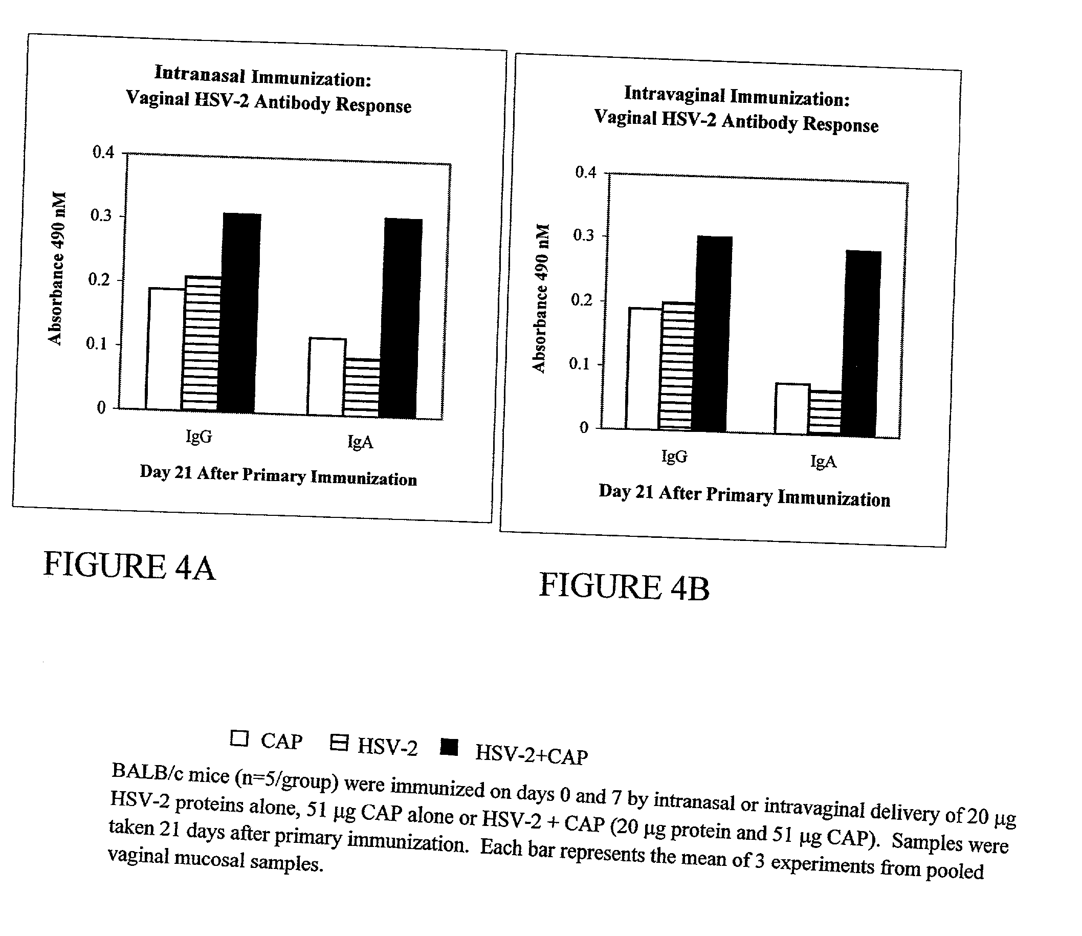
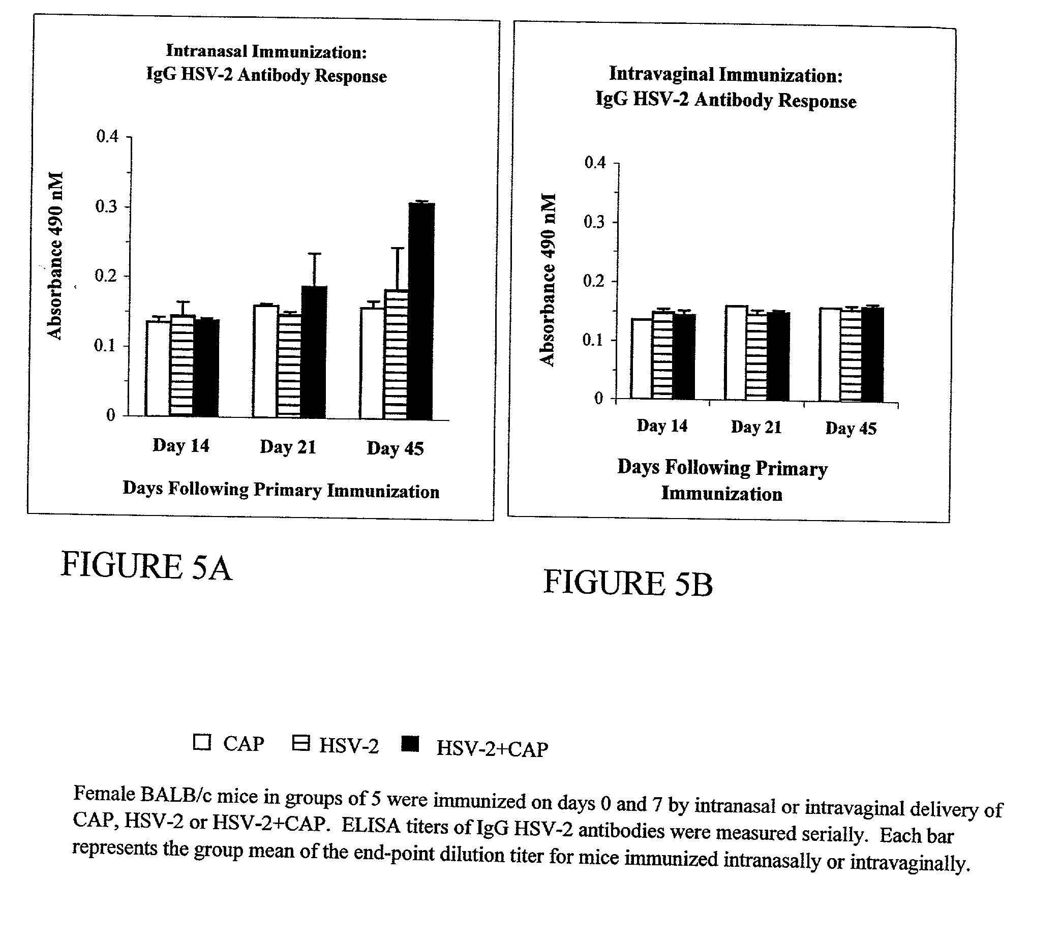
[0118] The HSV-2 protein of Example 2 was added to 75 ml or 12.5 mM calcium chloride, followed by the addition of 75 ml of 12.5 mM dibasic sodium phosphate and 15 ml of 15.6 mM sodium citrate similar to the particle formation methods described in Example 1. The solution was stirred until the final average particle size was less than 1,200 nm, as determined with a Coulter N4Plus Submicron Particle Sizer. The particle mixture containing entrapped HSV-2 protein was treated with cellobiose overnight and mixed again with 600 .mu.g HSV-2 protein for 1 hour at 4.degree. C. After washing off unbounded proteins with PBS, the HSV2-2+CAP vaccine formulation was ready for use.
PUM
| Property | Measurement | Unit |
|---|---|---|
| Diameter | aaaaa | aaaaa |
| Diameter | aaaaa | aaaaa |
| Pharmaceutically acceptable | aaaaa | aaaaa |
Abstract
Description
Claims
Application Information