Surface plasmon resonance imaging of micro-arrays

a surface plasmon and microarray technology, applied in the direction of material analysis, sequential/parallax process reactions, library screening, etc., can solve the problems of no imaging capability of "biacore" instruments, severely restricting the number of dna sequences that can be screened in a single experiment, and commercially available devices such as "biacore" instruments, which do not offer imaging capabilities

Inactive Publication Date: 2003-01-23
WISCONSIN ALUMNI RES FOUND
View PDF10 Cites 112 Cited by
  • Summary
  • Abstract
  • Description
  • Claims
  • Application Information

AI Technical Summary

Problems solved by technology

Although powerful, the "BIAcore" instrument has no imaging capabilities.
This severely limits the number of DNA sequences that can be screened in a single experiment.
However, as noted above, commercially-available devices, such as the "BIAcore" instrument, offer no imaging capabilities.
To use SPR imaging techniques, it is essential that the nucleic acid array be constructed on a noble metal surface, and for this reason DNA arrays on glass supports from commercially available sources such as Affymetrix (Santa Clara, Calif.) are not a viable option.
However, the application of such technology for quantitative measurement of biomolecules has been limited by the high costs and laborious techniques associated with radioactive and fluorescent labeling and detection (Lockhart et al.
However the current techniques remain impractical for large-scale screening of biomolecules, primarily because they require photo-patterning or reversible protection chemistry, which make these techniques time-consuming and often prohibitively expensive.
Current micro-array techniques also require large amou

Method used


Image

  • Surface plasmon resonance imaging of micro-arrays
  • Surface plasmon resonance imaging of micro-arrays
  • Surface plasmon resonance imaging of micro-arrays

Examples


example 1

Demonstration of 1-Dimensional Array for Detection Nucleic Acids

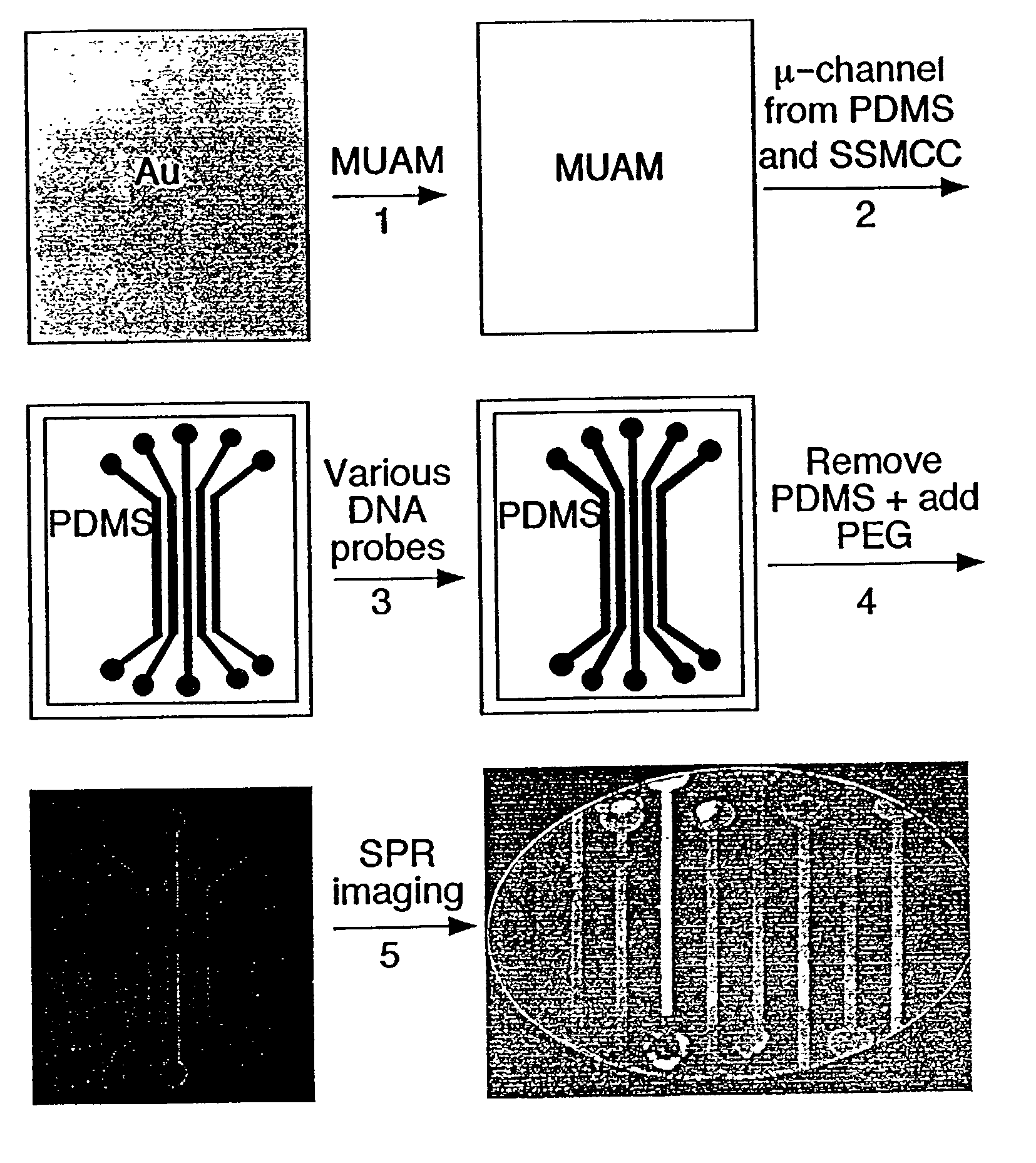
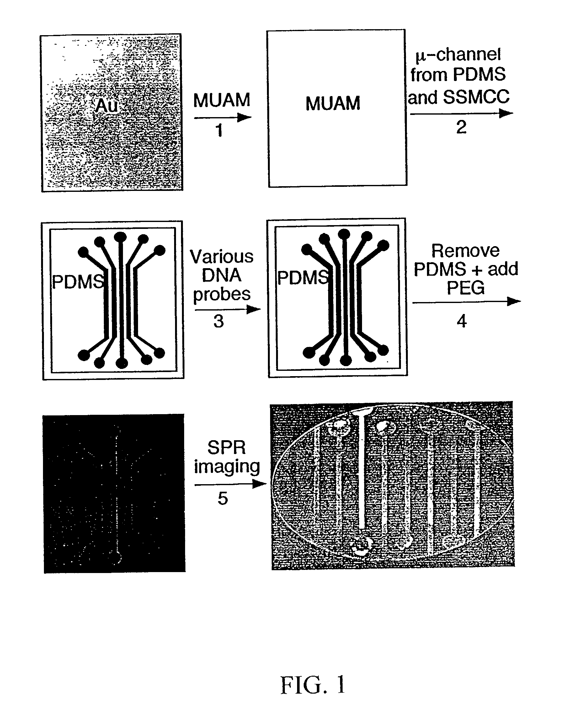
[0091] Referring now to FIG. 1, which shows the fabrication methodology according to the present invention for creating nucleic acid micro-arrays through the use of micro-fluidic channels in PDMS:

[0092] (1) A gold thin film surface is reacted with an amine-terminated alkanethiol (11-mercaptoundecylamine, MUAM) from a 1 mM ethanolic solution for two hours in order to form a self-assembled monolayer on the gold surface. (2) PDMS micro-channels were fabricated using the previously-described 1:1 photolithography technique of Anderson et al. (2000), and then attached to the MUAM-modified gold surface. A surface pattern is created by flowing a heterobifunctional linker, sulfosuccinimidyl 4-(N-maleimidomethyl)cyclohexane-1-carboxylate(SSMCC), through the PDMS micro-channels over the gold surface. The SSMCC reacts with the MUAM to create a maleimide-terminated alkanethiol monolayer. In order to overcome insufficient flow by cap...

example 2

Fabrication of 2-Dimensional DNA Micro-arrays Using Micro-channels

[0098] Referring now to FIG. 3A, in this example, the use of micro-fluidic channels has been extended to create 2-dimensional arrays based on the 1-dimensional DNA micro-arrays described in example 1. In these experiments, a 1-dimensional array as described in example 1 is first created on the gold surface. Then, a second set of PDMS micro-channels is attached to the surface so as to intersect the previously-deposited 1-dimensional array. This creates a 2-dimensional array of intersections that can be used to detect adsorption onto the surface-bound nucleic acids using either fluorescence microscopy or SPR imaging. Target solutions of complementary DNA or RNA are delivered via the second set of micro-channels so that the target solutions pass over the nucleic acids bound to the surface. The target solutions are allowed to contact the bound nucleic acids for a time sufficient to allow binding of the target to the bound...

example 3

SPR Imaging Using 2-Dimensional Arrays

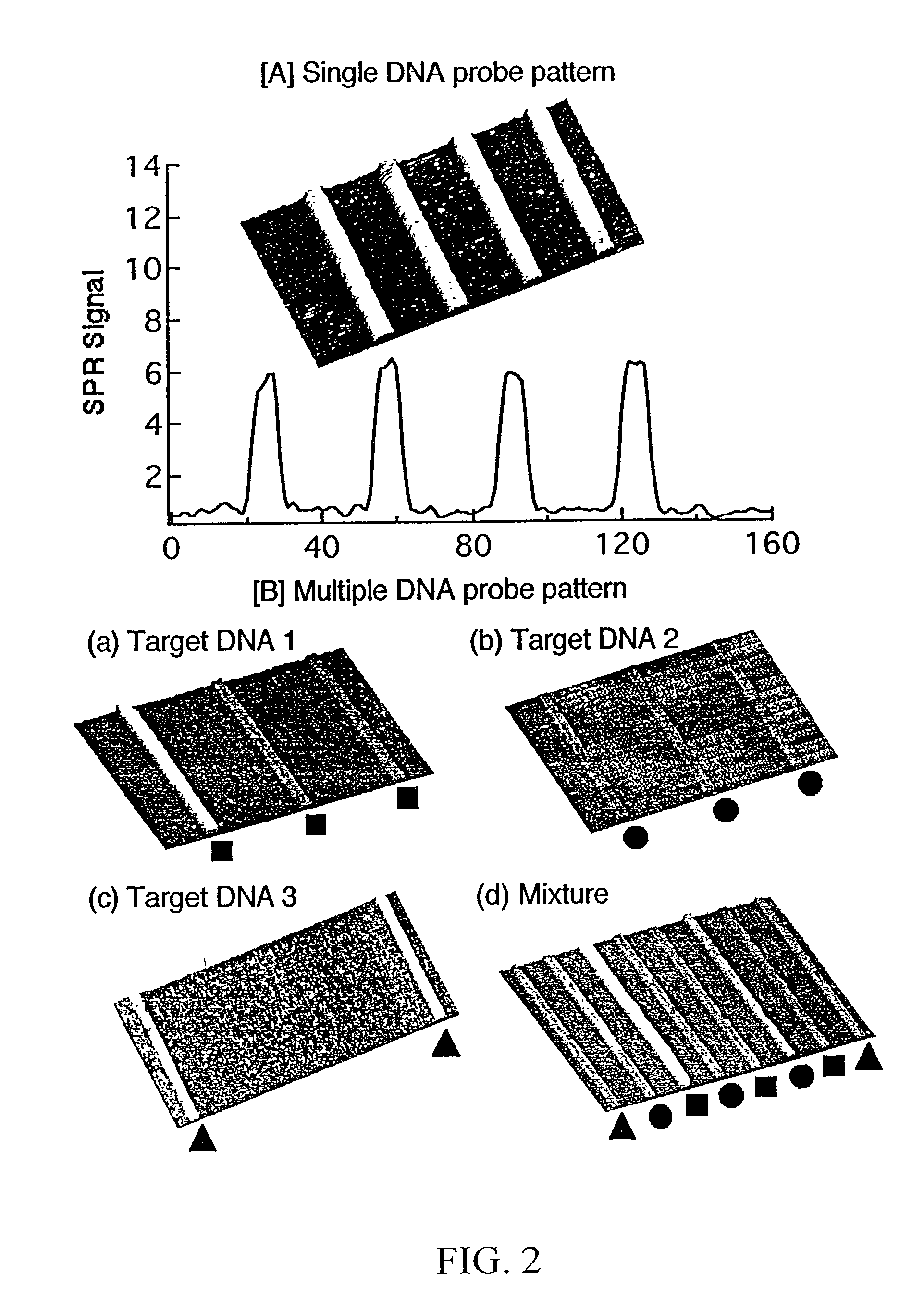
[0101] Referring now to FIGS. 4A through 4D, these figures are SPR difference images of specific hybridization of complementary DNA onto a 2-dimensional DNA array. Each array element is 300 .mu.m by 300 .mu.m. Specific hybridization and adsorption of each probe was achieved without nonspecific adsorption or other interferences. Of particular note is that these DNA micro-arrays reduce the sample volume required for SPR analysis by an order of magnitude when compared to conventional flow cell system.

[0102] In this example, three different ssDNA probes were affixed to a gold surface in roughly parallel channels, one type of DNA per channel using the exact approach described in example 1. A PDMS micro-channel array having two parallel channels was then affixed on top of, and perpendicular to, the previously-deposited ssDNA probes, so that each channel intersected all three of the previously-deposited ssDNA probes. Complementary ssDNA was then flowed...


PUM

PropertyMeasurementUnit
Volumeaaaaaaaaaa

Abstract

Disclosed is a method for fabricating 1-dimensional micro-arrays using parallel micro-fluidic channels on chemically-modified metal, carbon, silicon, and/or germanium surfaces; a muL detection volume method that uses 2-dimensional nucleic acid micro-arrays formed by employing the 1-dimensional DNA micro-arrays in conjunction with a second set of parallel micro-fluidic channels for solution delivery, and the 1-dimensional and 2-dimensional arrays used in the methods. The methodology allows the rapid creation of 1- and 2-dimensional arrays for SPR imaging and fluorescence imaging of DNA-DNA, DNA-RNA, DNA-protein, and protein-protein binding events. The invention enables very small volumes necessary for a variety of bioassay applications to be analyzed by SPR. Target solution volumes as small as 200 pL can be analyzed.

Description

PRIORITY[0001] Priority is hereby claimed to provisional application Ser. No. 60 / 304,246, filed Jul. 10, 2001, the entire contents of which is incorporated herein by reference.[0003] The invention is directed to a method of producing chemical micro-arrays (preferably nucleic acid micro-arrays) on chemically-modified gold and silicon films using micro-fluidics techniques, the micro-arrays produced thereby, and a method of analyzing the arrays using surface plasmon resonance (SPR) techniques. The SPR imaging techniques can, for example, be used to detect the affinity binding of unlabeled biological molecules onto arrays attached to the gold surface. The inventive techniques described herein provide simple, inexpensive, and powerful tools for the label-free detection of biomolecules.INCORPORATION BY REFERENCE[0004] All of the documents cited herein are incorporated by reference.DESCRIPTION OF THE RELATED ART[0005] The binding of proteins to DNA plays a pivotal role in the regulation an...

Claims


Application Information

Patent Timeline
IPC IPC(8): B01J19/00B01L3/00C12Q1/68C12Q1/6837G01N21/55
CPCB01J19/0046B01J2219/00274B01L3/502707B01L2200/12B01L2200/16B01L2300/0636B82Y15/00B82Y30/00C12Q1/6837G01N21/253G01N21/553G01N21/6458C12Q2565/629
Inventor CORN, ROBERT M.LEE, HYE JINGOODRICH, TERRY T.
Owner WISCONSIN ALUMNI RES FOUND
Why Eureka
Try Eureka