
Established cell line of microglia

a cell line and established technology, applied in the field of subcultivation established microglia, can solve the problems of difficult supplementary therapy, no other method than direct injection of the substance, and specific introduction into the brain, and achieve the effect of easy monitoring

Inactive Publication Date: 2004-04-15
JAPAN SCI & TECH CORP
View PDF0 Cites 2 Cited by
  • Summary
  • Abstract
  • Description
  • Claims
  • Application Information

AI Technical Summary

Benefits of technology

[0033] The established cell line of microglia of the present invention can be purified from mouse or rat brain cells after primary culture and then separated by the following means from this purified microglia. In addition, because the established cell line of microglia of the present invention is easily handled and has affinity for the brain, a gene or drug can be introduced into the established cell line of microglia and injected into peripheral blood vessels to express the gene in the brain or to deliver the drug to the brain specifically.
[0040] The adherent cells which do not float by the above mechanical stimulation are treated with trypsin-EDTA, then divided into single cells, plated on a non-treated plastic dish (non-coat plastic vessel), and allowed to adhere to it. Generally, a conventional culture vessel is treated with chemicals so as to be positively charged, but a dish not subjected to this treatment should be used in order to obtain the adherent cells. After incubation in a CO.sub.2 incubator at 37.degree. C. for 1 hour, cells floating in the medium after mechanical stimulation are removed, and cells capable of proliferation on the vessel are recovered by Rubber Policeman, etc., and the same procedure is repeated twice to obtain cells to be used in establishing a cell line of microglia. Although the purified microglia thus obtained is of adequate purity, cell sorter, etc. can also be used to further improve purity.
[0054] As a means of efficiently introducing a gene into the established cell line of microglia, there is for example a method of using DOTAP (Boehringer-Mannheim Co.). That is, there is a method of culturing the established cell line of microglia together with a desired gene in a gene-introducing medium containing DOTAP. They are cultured in a CO.sub.2 incubator at 37.degree. C. for 16 to 24 hours and further cultured together with rGM-CSF for 30 to 72 hours (preferably for 48 hours) in a medium for culturing microglia.
[0057] When this screening method is applied to the microglia, the microglia coming to express the introduced gene stably and constantly recognize dead cells thus expressing a strong phagocytic ability and simultaneously undergoing activation to lose their cell growth ability. Accordingly, the method of introducing a gene in the present invention is preferably a method wherein an expression vector modified so as to be capable of expressing a fluorescent protein, preferably green florescent protein (GFP) derived from an aurelia, in higher animals is used in place of a drug resistance gene, and by using a difference in fluorescence intensity, the microglia is separated as the desired cells exhibiting stable and constant expression.
[0059] The cells can also be confirmed using magnetic resonance image (MRI), positron emission tomography (PET), etc. For example, contrast media, etc. for MRI may be incorporated into the cells which are then injected into an animal so that the cells can be monitored in the animal. According to these methods, it is not necessary to kill the animal, and the cells can be monitored easily in a non-invasive manner.

Problems solved by technology

In the brain, however, the blood-brain barrier is present, so it is difficult to conduct supplementary therapy and to introduce an effective drug, and even if a substance (e.g. anticancer drug, DNA, etc.) is introduced from a peripheral position, it cannot be introduced specifically into the brain.
Therefore, there was no method other than direct injection of the substance by surgical operations.
However, even in this method too, incorporation of liposomes into the brain is as low as about 1% based on the injection amount, so this method cannot be said to be specific for the brain.
As described above, the blood-brain barrier is present in the brain, so the brain is almost free of infiltration with cells or a substance from the periphery, thus making it difficult to introduce a drug or a gene into the brain.
For this primary culture, however, the brain should be excised and purified for use, and primary culture usually requires a period of about 2 weeks, so the procedures are cumbersome.
Further, the cells are difficult to proliferate during culture and hard to subculture, so after primary culture, it is extremely difficult to introduce a gene into the microglia to express it therein.

Method used


Image

  • Established cell line of microglia
  • Established cell line of microglia
  • Established cell line of microglia

Examples


example 1

Separation of Established Clone of Microglia

[0061] (1) Isolation of Microglia

[0062] Brains were excised from newborn mice (C57BL6, op / op) and newborn rats (Fisher), and meninges were removed in an ice-cold microglia culture medium (referred to as Mi medium; Eagle's MEM containing 10% bovine serum, 0.2% glucose and 5 .mu.g / ml bovine insulin). The cells of meninges were divided into single cells with a Pasteur pipette or nylon mesh and then cultured in Mi medium. For the cells from the mouse brains, 20 ml Mi medium was used per one brain, and for the cells from the rat brains, 40 ml Mi medium was used per one brain, and the former cells were incubated in 2 culture vessels of 10 cm diameter and the latter cells in 4 culture vessels of 10 cm diameter in a CO.sub.2 incubator (5% CO.sub.2, 95% air) at 37.degree. C. for 10 to 14 days. The medium was exchanged with fresh one every 3 to 4 days.

[0063] When phase-bright round cells (PBRCS) appeared, the PBRCs were removed by mechanical shaking...

example 2

Introduction of a Gene into the Microglia of the Present Invention

[0088] Vector ptk.beta.(Clonetech Co.) for expression of lac Z gene derived from E. coli and DOTAP lipid (Boehringer-Mannheim Co.) were mixed to be a final concentration of 1 .mu.g / ml. The mixture was mixed with a serum-containing medium, then added to the established cell line of microglia of the present invention and treated for 16 hours. As the control, the established microglia of the invention into which the gene was not introduced, and macrophages obtained in the same manner as in Example 1, were used.

[0089] Then, the cells were further cultured for 48 hours in a usual medium (EMEM plus 10% FCS) and then stained with the fluorescent pigment as described in Example 1 (b-1) in order to examine whether the gene was delivered to and expressed in the brain, as follows: An artery in the left armpit of a mature rat (250 to 300 g) under anesthesia with Nembutal was exposed. After hemostatic treatment, a cannula was inse...

example 3

Introduction of Chemical Substance into the Microglia of the Invention, and Specific Introduction Thereof into the Brain

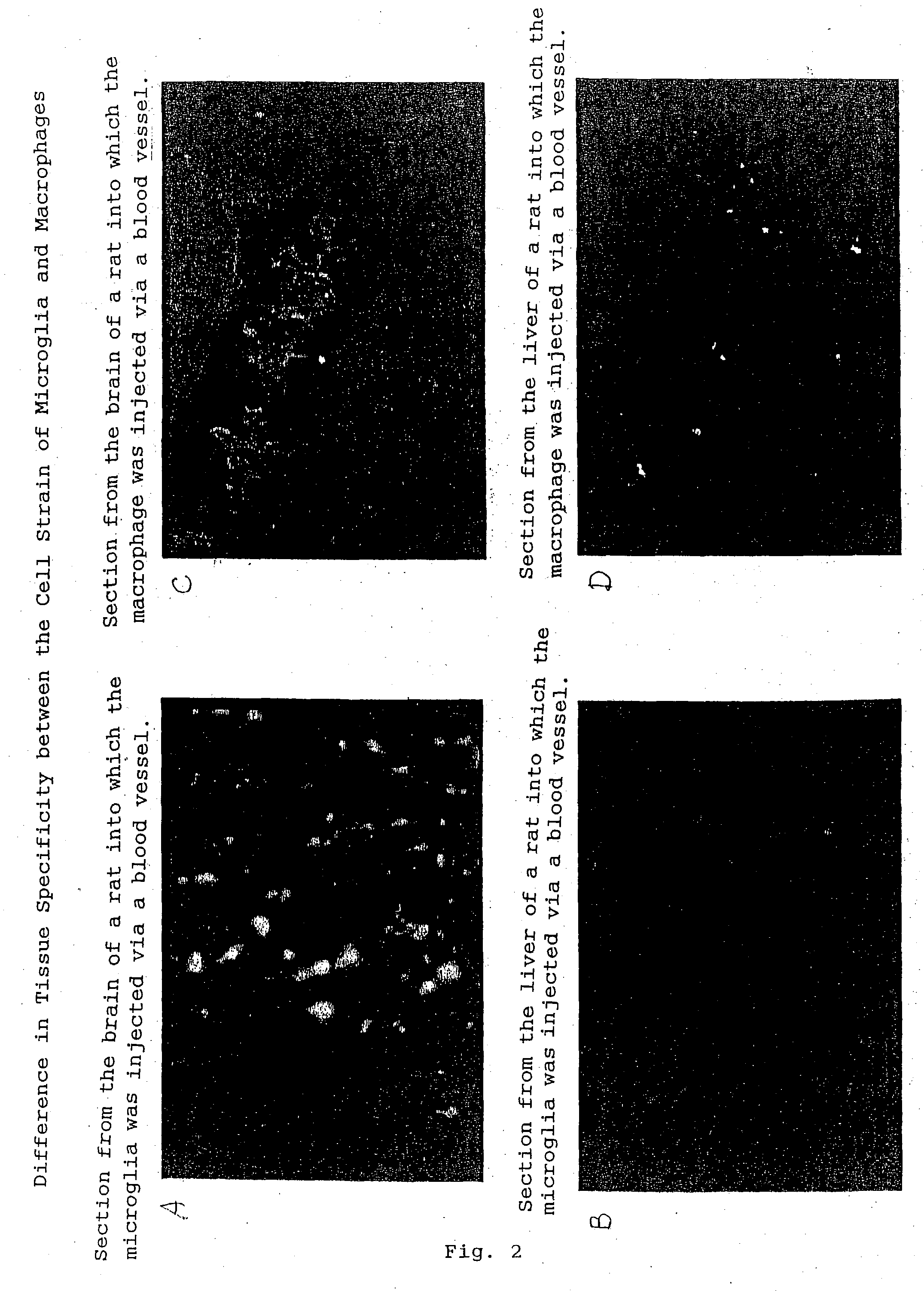
[0093] The fluorescent pigment PKH26 used in Example 1 forms granules in diluent B. The microglia cell strain of the present invention incorporates these granules specifically and transfers them to the brain, so the fluorescent pigment PKH26 was used as a model of chemical substance (anti-tumor drug).

[0094] As a result, when the established cell line of microglia of the present invention was introduced, many fluorescent cells were observed in normal brain cells but not observed in the liver. It therefore follows that the microglia of the present invention transfers the chemical substance (drug) into the brain specifically.


PUM

PropertyMeasurementUnit
Fluorescenceaaaaaaaaaa

Abstract

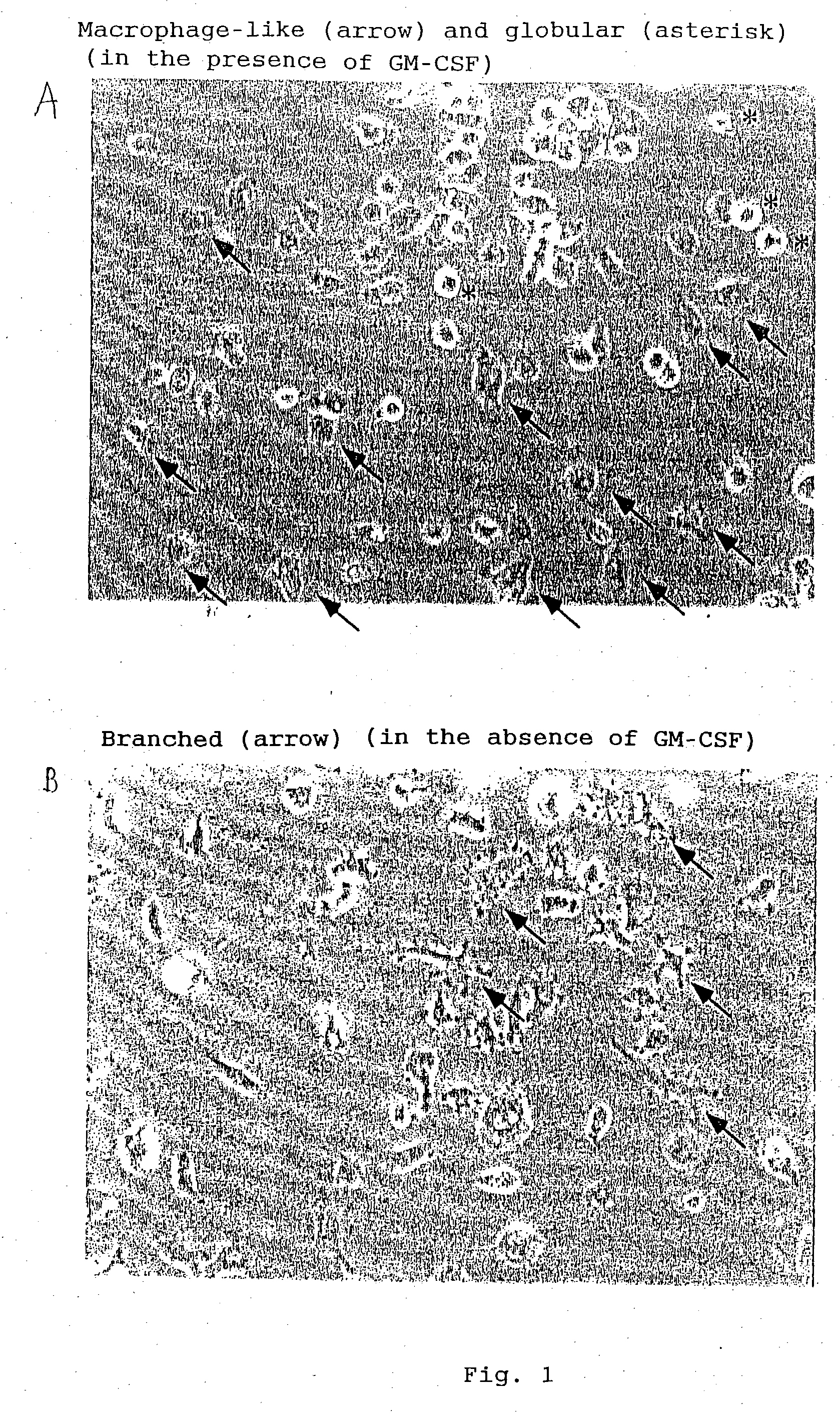
A subcultivatable, established microglia having the following properties. (a) Form: Both or either of a macrophage-like or spherical form in the presence of a granulocyte-macrophage colony stimulation factor and a branched form similar to branched microglia present in the brain in the absence of the factor. (b) Functional characteristics: specific affinity for the brain highly poor phagocytic action. (c) Cell proliferation: proliferative depending upon a granulocyte-macrophage colony stimulation factor. Preparation, separation, and screening methods of the microglia, a pharmaceutical composition using the microglia, and a method for treatment of cerebral diseases using the composition.

Description

[0001] The present invention relates to a subcultivatable, established cell line of microglia, a method of separating the same, and use thereof as a pharmaceutical carrier.[0002] Further, the present invention relates to a microglia into which an extraneous gene or a drug was introduced, a method of introducing it, and a pharmaceutical composition comprising the same.[0003] There are quite a number of hereditary diseases in the nervous system, which occur due to various causative factors, for example by defect of a single enzyme, etc. or by unknown reasons. Under these circumstances, supplementary therapy is used to cope with a large number of such diseases.[0004] A large number of studies have been made worldwide on the system of selective delivery to the brain. For introducing a gene into the brain in an animal, a method of using neuronphilic viruses as adenovirus vectors is devised, and a system for introducing a gene specifically into neurons is known (Kozarsky, K. F and Wilson,...

Claims


Application Information

Patent Timeline
IPC IPC(8): A61K35/30A61K38/00A61K48/00C12N5/07C12N5/0786C12N5/0787C12N5/079
CPCA61K38/00A61K48/00C12N2510/00C12N2501/22C12N5/0622A61P25/00
Inventor SAWADA, MAKOTO
Owner JAPAN SCI & TECH CORP
Learn More