Tissue scaffold having aligned fibrils, apparatus and method for producing the same, and artificial tissue and methods of use thereof

a tissue and fibril technology, applied in the field of tubular tissue scaffolds, can solve the problems of adverse tissue reactions, inflexible degradable polyesters, and the most difficult challenges, and achieve the effects of minimal immunological response, improved structural integrity, and sufficient structural strength

Active Publication Date: 2005-01-13
UNIVERSITY OF SOUTH CAROLINA
View PDF48 Cites 165 Cited by
  • Summary
  • Abstract
  • Description
  • Claims
  • Application Information

AI Technical Summary

Benefits of technology

[0025] Among the several advantages found to be achieved by the present invention, therefore, may be noted the provision of a tubular tissue scaffold having sufficient structural strength to withstand pressure; the provision of a scaffold having a composition that is biologically degradable in vivo and will result in a minimal immunological response from a host; the provision of a tissue scaffold having a structural composition that allows the penetration of cells and provides cells the requisite signals to develop an in vivo functional phenotype; the provision of an apparatus and a method for the production of such a tubular tissue scaffold, and; the provision of artificial tissue comprising cells attached to a tissue scaffold of biopolymer fibrils that is non-immunogenic, has a construction that mimics that of cardiac tissue, and has improved structural integrity.

Problems solved by technology

One of the most difficult challenges is directing the behavior of specialized cells outside of the body to mimic the normal, endogenous phenotype those cells exhibit in vivo.
However, those degradable polyesters tended to be inflexible, and their degradation in vivo has been associated with adverse tissue reactions.
However, some problems remain to be solved in the use of collagen as a scaffolding material for applications requiring structural and mechanical stability, such as for vascular prosthetics.
This is at least partly due to an inability to isolate collagen possessing the physical properties required to maintain necessary mechanical integrity of a scaffold, as it is remodeled in vivo, for use in, for example, cardiovascular indications.
Also, despite the advances in biomaterials research, and the elucidation of the molecular biology of cell behavior and cell:matrix interactions, the gap between in vitro engineered tissue and biologically functional implantable organs remains significant.

Method used


Image

  • Tissue scaffold having aligned fibrils, apparatus and method for producing the same, and artificial tissue and methods of use thereof
  • Tissue scaffold having aligned fibrils, apparatus and method for producing the same, and artificial tissue and methods of use thereof
  • Tissue scaffold having aligned fibrils, apparatus and method for producing the same, and artificial tissue and methods of use thereof

Examples


example 1

[0131] This example illustrates the preparation of a biopolymer gel comprising type I collagen from a bovine steer hide.

[0132] Collagen was prepared from the hide of an 18-month old bovine steer by removal of the superficial epidermis including the hair and follicle pits. To isolate the collagen the hide was cut into 4×6 cm strips and washed with three changes of deionized (DI) water for 1 hr. each (the second wash contained 0.2% NaHCO3). The remaining follicles and noncollagenous proteins were removed by bathing in a solution of 0.6% NaHCO3, 2% Ca(OH)2, and 4.3% NaHS for 30 min at 20° C. After three washings in DI water, the strips were treated overnight in 2% Ca(OH)2 at 4° C. The remaining fat and epidermis were trimmed from the strips the following day. The strips were then placed in a 2M NaCl solution and neutralized with HCl to a pH of 6.8-7.0. The strips were then washed three times in DI water, cut into 0.5×2 cm pieces, and placed in a solution of 0.5 N acetic acid with or w...

example 2

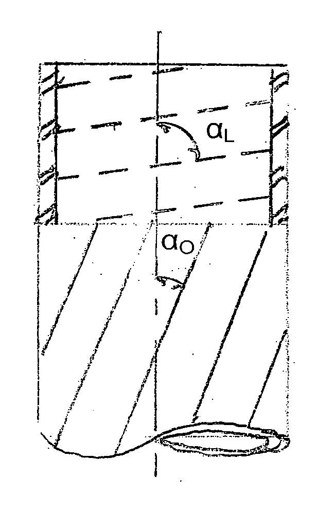
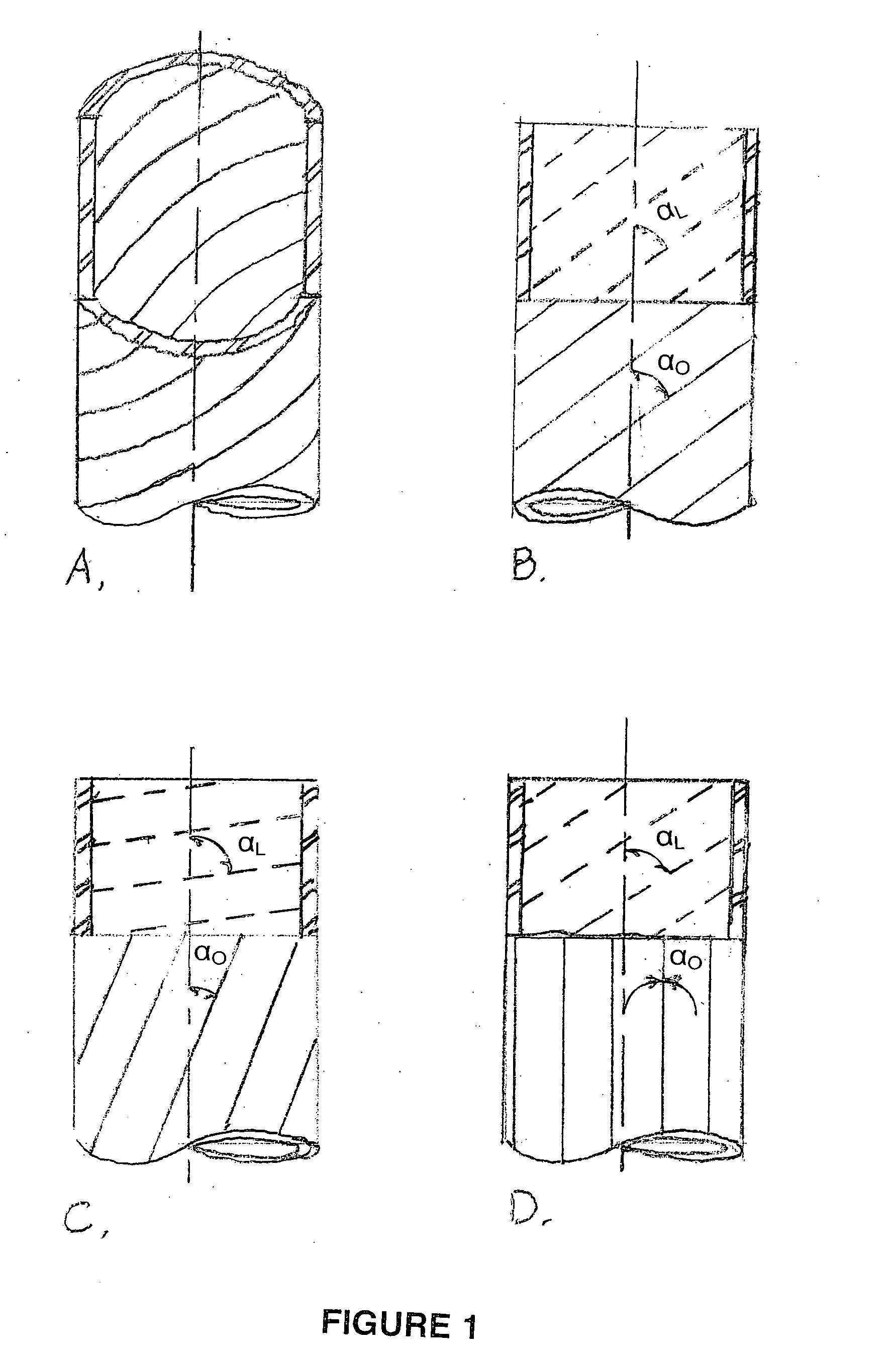
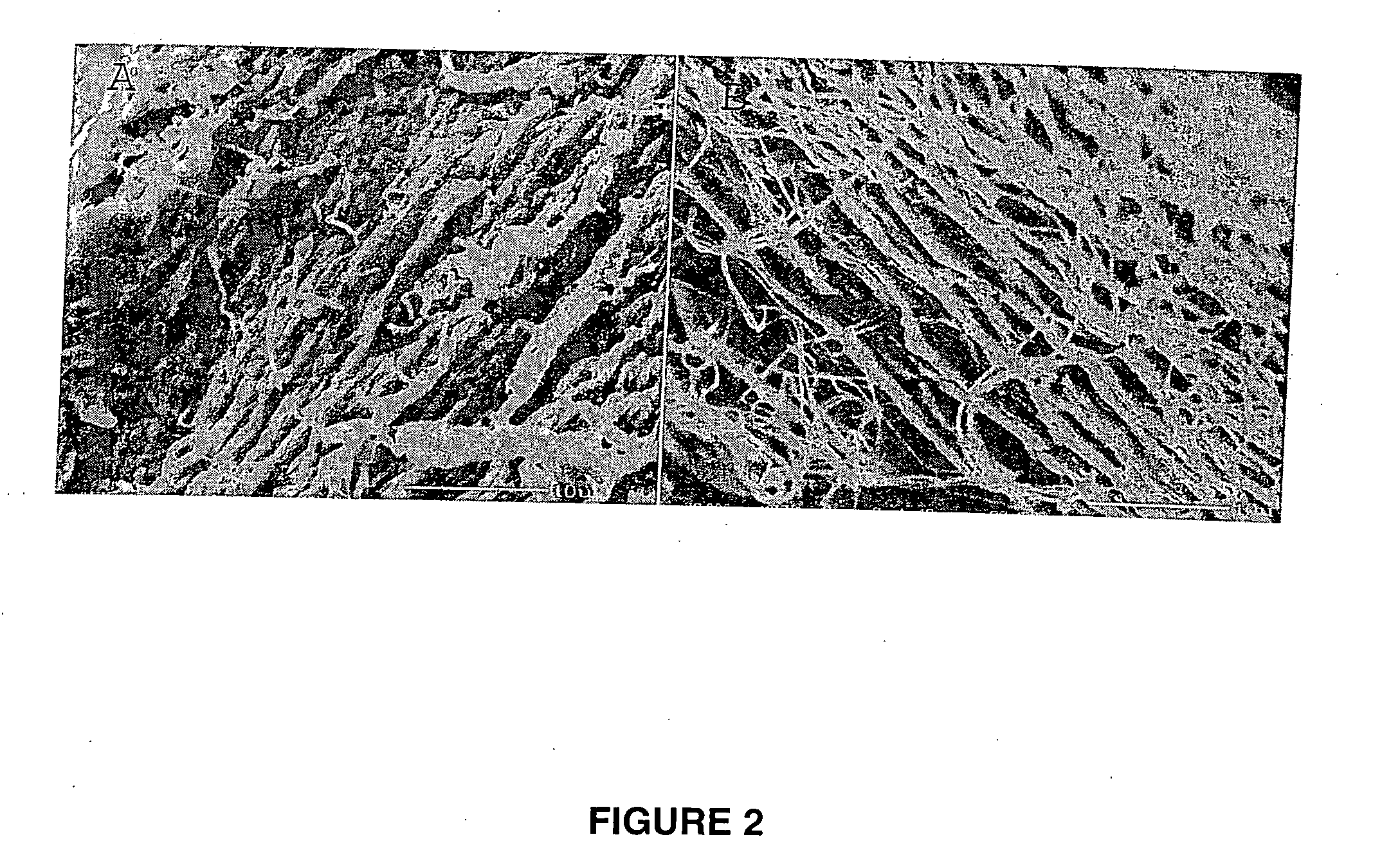
[0133] This example illustrates the production of a collagen tubular tissue scaffold having aligned fibrils by using a counter-rotating cone extruder.

[0134] Collagen tubes were produced by loading a collagen gel dispersion, produced as described in Example 1, into a 60 ml syringe that was placed into a syringe pump (having an adjustable feed rate of 0-20 cc / min) that fed the collagen into the feed port of a counter-rotating cone extruder that was fabricated from Delrin (Tri Star plastics, Reading, Pa.) according to the illustration shown in FIG. 1. The drive motor was a 90 volt 0.06 hp DC motor (Baldor Electric, Ft. Smith Ark.) with a regenerative feedback drive and a 5 kΩ potentiometer for user interface (KB electronics, Coral Springs Fla.). The motor is coupled to the pinion gear which drives the outer rotating member and the inner rotating cone drive gears. The direct drive system can control speeds from 0 to 1800 rpm with a 2:1 reduction from the pinion gear to the drive gears....

example 3

[0144] This example illustrates seeding of cardiac myocyte on a collagen tubular tissue scaffold.

[0145] Collagen tubes were produced as described in Example 2. Prior to the addition of myocytes, the collagen tubes were sectioned into 1.25 cm lengths. The small collagen tube sections were placed in sterile Mosconas solution (0.14M NaCl, 0.0027M KCl, 0.012M NaHCO3, 4.2×10⁻⁵ M NaH2PO4, 0.0094M glucose) and exposed to ultraviolet (UV) light for 4 hours to sterilize. Following UV treatment, fresh Marconas solution with 0.01 mg / ml gentamycin, 4 μg / ml Amphotericin-B, and 10 μg / ml fibronectin was added to a culture dish containing the collagen tube sections and incubated for 24 hours with 5% CO2 at 37° Celsius.

[0146] Isolation of neonatal cardiac myocytes was performed as described by Simpson et al., J Cell Physiol, 161:89-105 (1994). For seeding, collagen tube sections were placed in 150 mm culture dishes and 0.5 ml of a cellular suspension containing 2×10⁶ cells / ml was injected into the...


PUM

PropertyMeasurementUnit
diameteraaaaaaaaaa
diameteraaaaaaaaaa
diameteraaaaaaaaaa

Abstract

A tubular tissue scaffold is described which comprises a tube having a wall, wherein the wall includes biopolymer fibrils that are aligned in a helical pattern around the longitudinal axis of the tube where the pitch of the helical pattern changes with the radial position in the tube wall. The scaffold is capable of directing the morphological pattern of attached and growing cells to form a helical pattern around the tube walls. Additionally, an apparatus for producing such a tubular tissue scaffold is disclosed, the apparatus comprising a biopolymer gel dispersion feed pump that is operably connected to a tube-forming device having an exit port, where the tube-forming device is capable of producing a tube from the gel dispersion while providing an angular shear force across the wall of the tube, and a liquid bath located to receive the tubular tissue scaffold from the tube-forming device. A method for producing the tubular tissue scaffolds is also disclosed. Also, artificial tissue comprising living cells attached to a tubular tissue scaffold as described herein is disclosed. Methods for using the artificial tissue are also disclosed.

Description

CROSS REFERENCE TO RELATED APPLICATIONS [0001] This application is related to and claims the priority benefit of U.S. Provisional Application Ser. Nos. 60 / 475,680; 60 / 475,866; and 60 / 475,986, filed Jun. 4, 2003, each of which is incorporated herein by reference in its entirety.STATEMENT REGARDING FEDERALLY SPONSORED RESEARCH OR DEVELOPMENT [0002] The U.S. Government has a paid-up license in this invention and the right in limited circumstances to require the patent owner to license others on reasonable terms as provided for by the terms of grant no. 1 K25 HL67097 awarded by the U.S. National Institutes of Health of the Department of Health and Human Services.BACKGROUND OF THE INVENTION [0003] (1) Field of the Invention [0004] The present invention relates to a tubular tissue scaffold, and, in particular, to a tubular tissue scaffold having aligned biopolymer fibrils, for use in tissue engineering applications; an apparatus and method of producing a tubular tissue scaffold having ali...

Claims


Application Information

Patent Timeline
Patent Type & Authority Applications(United States)
IPC IPC(8): A61F2/02A61F2/04A61F2/06A61K38/17B29C48/09B29C48/32B29C48/33C12M3/00C12NC12N5/08C12N11/02
CPCB29C47/0023C12M25/14B29C47/24C12M21/08B29C48/09B29C48/33
Inventor YOST, MICHAEL J.GORE, C. MICHAELTERRACIO, LOUISGOODWIN, RICHARD L.GOLDSMITH, EDIE C.
Owner UNIVERSITY OF SOUTH CAROLINA
Why Eureka
Try Eureka