
Methods of identifying kinetically stable proteins

a kinetic stability and protein technology, applied in the direction of fluid pressure measurement, liquid/fluent solid measurement, peptide measurement, etc., can solve the problems of incomplete protein protection, no structural consensus, and poorly understood physical basis of kinetic stability, so as to achieve fast and efficient means, fast and efficient results, and slow unfolding rate

Inactive Publication Date: 2005-03-31
RENESSELAER POLYTECHNIC INST
View PDF7 Cites 2 Cited by
  • Summary
  • Abstract
  • Description
  • Claims
  • Application Information

AI Technical Summary

Benefits of technology

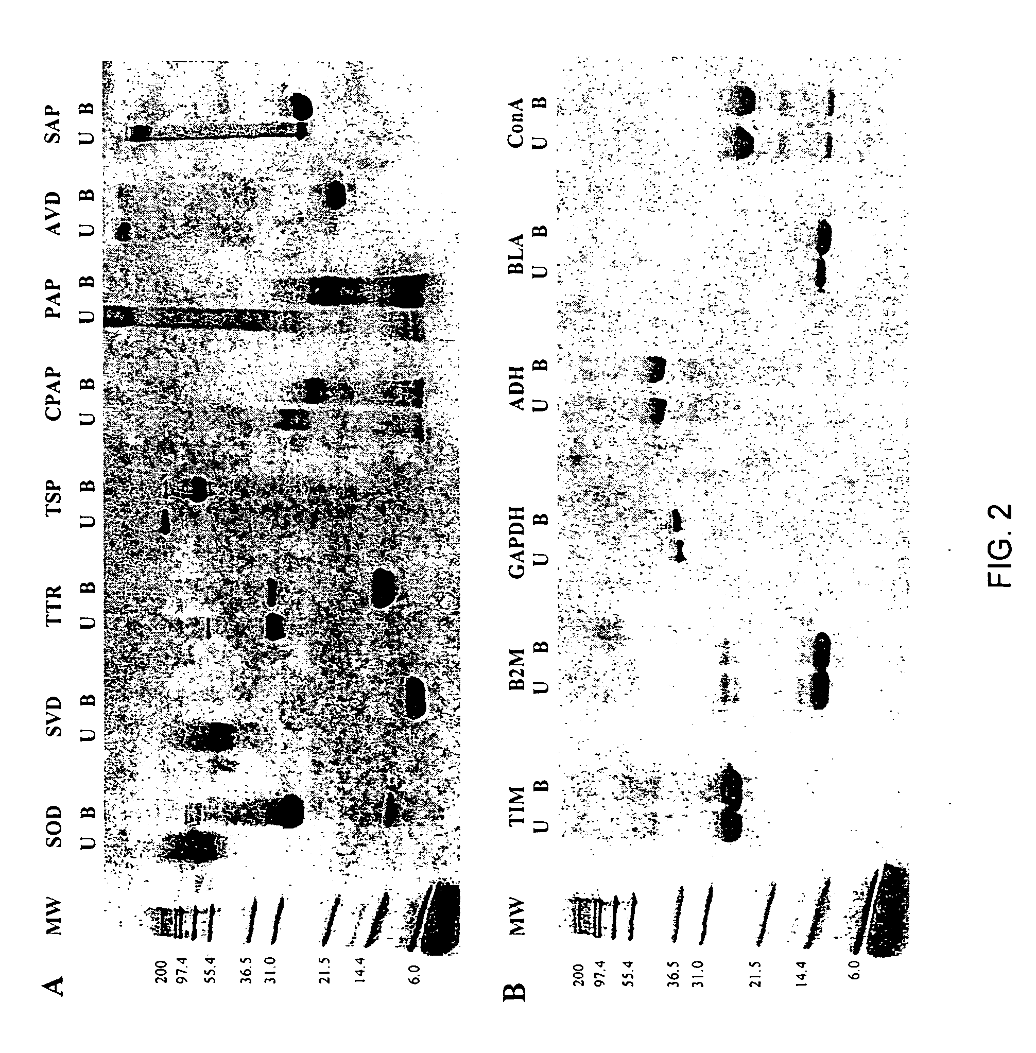
The present invention provides a fast and efficient means for identifying kinetically stable proteins. As used herein the term “kinetically stable protein” means a protein that is trapped in a specific conformation due to an unusually high unfolding barrier that results in very slow unfolding rates. The present inventors are the first to discover the existence of a correlation between kinetic stability and SDS-induced denaturation. Thus, the invention provides methods for identifying kinetically stable proteins comprising the step of testing the proteins for resistance to denaturation by SDS. In one embodiment, SDS-polyacrylamide gel electrophoresis (SDS-PAGE) is one simple method for quickly identifying and selecting kinetically stable proteins. This method has the advantage that proteins can be easily tested for kinetic stability without having to carry out unfolding experiments. Also, only microgram amounts of sample are needed, and the method is potentially suitable for identifying kinetically stable proteins present in cell extracts without need for purification. From an application perspective, this assay has the potential of being adaptable for various high-throughput applications to enhance the kinetic stability of proteins of interest. This could lead to proteins with greater shelf life and / or decreased tendency to aggregate, consistent with the suggestion that the deterioration of an energy barrier between native and pathogenic states as a result of mutation may be a key factor in the misfolding and aggregation of some proteins linked to amyloid diseases (4, 18). Such protein misfolding diseases include but are not limited to Alzheimer's disease, Parkinsons, prior-related encephalopathies, amyotrophic lateral sclerosis, and type II diabetes.

Problems solved by technology

In addition, thermodynamic stability alone does not fully protect proteins that are susceptible to irreversible denaturation and aggregation arising from partially denatured states that become transiently populated under physiological conditions (4).
The physical basis for kinetic stability is poorly understood and no structural consensus has been found to explain this phenomenon.

Method used


Image

  • Methods of identifying kinetically stable proteins
  • Methods of identifying kinetically stable proteins
  • Methods of identifying kinetically stable proteins

Examples


example 1

SDS-polyacrylamide Gel Eectrophoresis (SDS-PAGE) Assay. Lyophilized proteins were obtained from Sigma (papain (PAP), chymopapain (CPAP), avidin (AVD), and superoxide dismutase (SOD)) and Calbiochem (streptavidin (SVD), serum amyloid P (SAP), and transthyretin (TTR)). Salmonella phage P22 tailspike (TSP) protein was a gift from J. King (MIT). All the remaining proteins (Table 2) were obtained from Sigma with the exception of catalase, which was purchased from Calbiochem. Stock solutions (1 mg / mL) of all proteins studied except papain, chymopapain, and P22 tailspike protein were made using 10 mM sodium phosphate buffer (pH 7.0) (PB). Stock solutions (1 mg / mL) of papain and chymopapain were prepared with 25 mM 2-amino-2-hydroxymethyl-1,3-propanediol (Tris), 1 mM EDTA (pH 5.3). The stock solution of the P22 tailspike protein was 0.8 mg / mL in 50 mM Tris, 2 mM EDTA (pH 7.6). All electrophoresis samples contained ˜5 μg protein and 1% sodium dodecyl sulfate (SDS) in 0.125 M Tris (pH 6.8). ...

example 2

Proteolysis. For limited proteolysis experiments the concentration of the proteins was determined by weighing the lyophilized protein on a microbalance. Each proteolysis reaction mixture contained about 0.5 mg / mL protein and 5 μg / mL proteinase K (Fisher Scientific) in 25 mM Tris, 1μM EDTA (pH 8.3), and was incubated at 25° C. for 48 hours. The reaction was stopped with a solution of 2.5 μM phenylmethylsulfonyl fluoride, 4% SDS in 0.125 M Tris, 3.4 μM 1,4-Dithio-DL-threitol (pH 6.8)). Samples were boiled and gel electrophoresis was performed using 16% acrylamide Novex precast gels (Invitrogen). Running buffer was 0.1% SDS in Tris / Tricine buffer (pH 8.1).

example 3

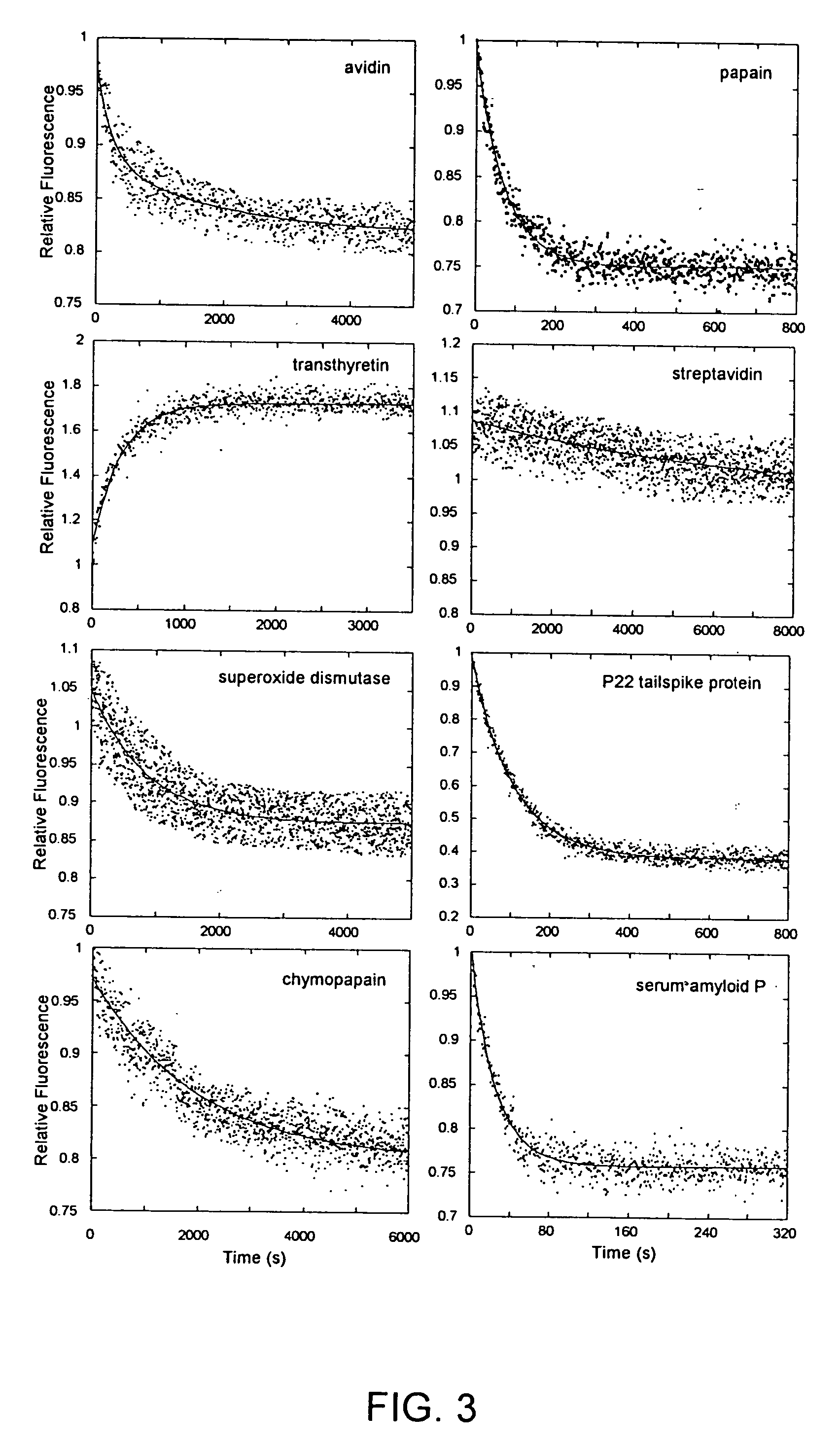
Fluorescence. Unfolding kinetics induced by guanidine hydrochloride (GdnHCl) were monitored using an F-4500 fluorescence spectrophotometer (Hitachi, Danbury, Conn.). The concentration of GdnHCl was determined using an Abbe Mark II refractometer (Leica, Buffalo, N.Y.). Protein solutions (0.05 mg / mL) in 25 mM PB, 0.20 M sodium chloride (pH 7.2) were treated with GdnHCl solution made using the same buffer to a final concentration of 6.6 M. The excitation / emission wavelengths used were: 275 / 350 nm (B2M), 275 / 360 nm (BLA, ConA, GAPDH, TIM), 280 / 320 nm (ADH), 295 / 360 nm (CPAP, TTR), 295 / 350 nm (PAP), 295 / 340 nm (SAP, TSP), 280 / 330 nm (SOD), 295 / 333 nm (SVD), and 280 / 340 nm (AVD). Kinetic traces were analyzed by fitting to a sum of exponentials.


PUM

PropertyMeasurementUnit
Electrical resistanceaaaaaaaaaa
Stabilityaaaaaaaaaa
Kinetic theoryaaaaaaaaaa

Abstract

The present invention provides a fast and efficient means for identifying kinetically stable proteins. As used herein the term “kinetically stable protein” means a protein that is trapped in a specific conformation due to an unusually high unfolding barrier that results in very slow unfolding rates. The present inventors are the first to discover the existence of a correlation between kinetic stability and SDS-induced denaturation. Thus, the invention provides methods for identifying kinetically stable proteins comprising the step of testing the proteins for resistance to denaturation by SDS. In one embodiment, SDS-polyacrylamide gel electrophoresis (SDS-PAGE) is one simple method for quickly identifying and selecting kinetically stable proteins.

Description

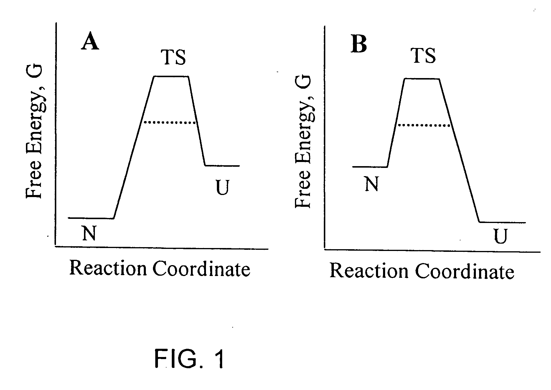
BACKGROUND OF THE INVENTION Kinetic stability is a poorly understood property of a select group of naturally occurring proteins that are trapped in their native conformations by an energy barrier, and consequently are resistant to unfolding. Kinetic stability can be best explained by illustrating the unfolding process as a simple equilibrium reaction between two protein conformations, the native folded state (N) and the unfolded state (U), separated by a higher energy transition state (TS) (FIG. 1). Since the height of the TS barrier determines the rate of folding and unfolding, kinetically stable proteins possess an unusually high energy TS, which results in extremely slow unfolding rates that virtually trap the protein in its native state (FIG. 1A). Even though the overall change in Gibbs free energy (ΔG) may be favorable for unfolding under extreme solvent conditions, such as high concentrations of denaturant, the high activation energy of the TS significantly slows down the unf...

Claims


Application Information

Patent Timeline
IPC IPC(8): G01N33/68
CPCG01N2500/00G01N33/6803
Inventor COLON, WILFREDOMANNING, MARTA
Owner RENESSELAER POLYTECHNIC INST
Learn More